. 2020 Jun 18;18(6):e3000679.
doi: 10.1371/journal.pbio.3000679. eCollection 2020 Jun.

Acute inhibition of centriolar satellite function and positioning reveals their functions at the primary cilium

Affiliations

Acute inhibition of centriolar satellite function and positioning reveals their functions at the primary cilium

Özge Z Aydin et al. PLoS Biol. .

Abstract

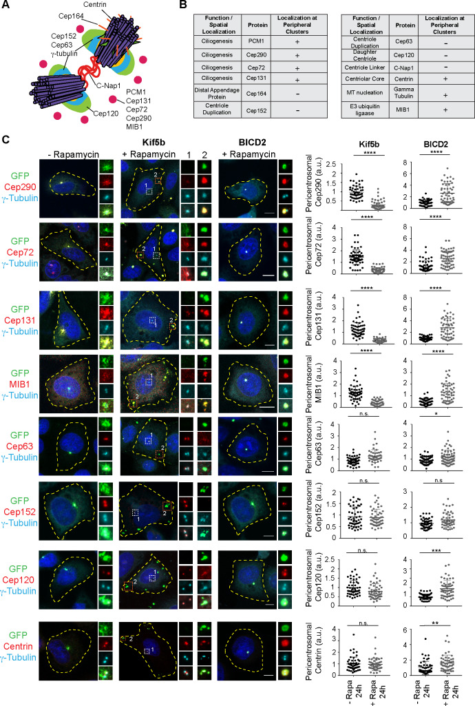
Centriolar satellites are dynamic, membraneless granules composed of over 200 proteins. They store, modify, and traffic centrosome and primary cilium proteins, and help to regulate both the biogenesis and some functions of centrosomes and cilium. In most cell types, satellites cluster around the perinuclear centrosome, but their integrity and cellular distribution are dynamically remodeled in response to different stimuli, such as cell cycle cues. Dissecting the specific and temporal functions and mechanisms of satellites and how these are influenced by their cellular positioning and dynamics has been challenging using genetic approaches, particularly in ciliated and proliferating cells. To address this, we developed a chemical-based trafficking assay to rapidly and efficiently redistribute satellites to either the cell periphery or center, and fuse them into stable clusters in a temporally controlled way. Induced satellite clustering at either the periphery or center resulted in antagonistic changes in the pericentrosomal levels of a subset of proteins, revealing a direct and selective role for their positioning in protein targeting and sequestration. Systematic analysis of the interactome of peripheral satellite clusters revealed enrichment of proteins implicated in cilium biogenesis and mitosis. Importantly, induction of peripheral satellite targeting in ciliated cells revealed a function for satellites not just for efficient cilium assembly but also in the maintenance of steady-state cilia and in cilia disassembly by regulating the structural integrity of the ciliary axoneme. Finally, perturbing satellite distribution and dynamics inhibited their mitotic dissolution, and mitotic progression was perturbed only in cells with centrosomal satellite clustering. Collectively, our results for the first time showed a direct link between satellite functions and their pericentrosomal clustering, suggested new mechanisms underlying satellite functions during cilium assembly, and provided a new tool for probing temporal satellite functions in different contexts.


Conflict of interest statement

The authors have declared that no competing interests exist.

Figures

Fig 1
Fig 1. Inducible dimerization of PCM1 with constitutively active motor domains targets satellites to the cell periphery or center.
Development and validation of the inducible satellite trafficking assay. (A) Representation of satellite redistribution to cell periphery or center upon inducible dimerization of the satellite scaffolding protein PCM1 with the constitutively active plus and minus end–directed molecular motor proteins. (B) Design of the inducible satellite-trafficking assay. PCM1 was tagged with GFP at the N terminus and FKBP at the C terminus. Co-expression of GFP-PCM1-FKBP with HA-Kif5b (1–269 aa)-FRB and subsequent rapamycin induction targets satellites to the cell periphery, where microtubule plus ends are concentrated. Co-expression of GFP-PCM1-FKBP with HA-BICD2 (1–198 aa)-FRB and subsequent rapamycin induction targets satellites to the cell center, where microtubule minus ends are concentrated. (C, D) Representative images for satellite distribution in control and rapamycin-treated HeLa cells. Cells co-expressing GFP-PCM1-FKBP with HA-Kif5b-FRB or HA-BICD2-FRB were treated with rapamycin for 1 hour. Twenty-four hours after rapamycin induction, cells were fixed and stained with antibodies against GFP, PCM1, and gamma-tubulin. DNA was stained with DAPI. Cell edges are outlined. Scale bar, 5 μm. (E, F) Endogenous PCM1 exhibits the same localization pattern as GFP-PCM1-FKBP. Pericentrosomal PCM1 levels was quantified at 6 hours and 24 hours after rapamycin treatment of cells expressing GFP-PCM1-FKBP with (E) HA-Kif5b-FRB or (F) HA-BICD2-FRB. Average mean intensity of pericentrosomal levels in control cells were normalized to 1. n ≥ 25 cells per experiment. Data represent mean value from two experiments per condition, ±SD (****p < 0.0001). (G) Dynamics of satellite redistribution to the cell periphery or center upon rapamycin addition. Cells co-expressing GFP-PCM1-FKBP with HA-Kif5b-FRB or HA-BICD2-FRB were imaged before and after rapamycin addition using time-lapse microscopy. Imaging was performed more frequently (every 2 minutes) in BICD2-expressing cells relative to Kif5b-expressing cells (every 5 minutes). Representative still frames from time-lapse experiments were shown. Cell edges are outlined. Time, minutes after rapamycin addition. Scale bar, 5 μm. (H) Satellites concentrate around mother centriole in cells co-expressing GFP-PCM1-FKBP with HA-BICD2-FRB after rapamycin treatment. Cells were stained for GFP, mother centriole marker Cep164, and gamma-tubulin. DNA was stained with DAPI. Cell edges are outlined. Scale bar, 5 μm. (I) Peripheral and centrosomal satellite clusters were maintained upon microtubule depolymerization. Cells with peripheral or centrosomal satellite clusters were treated with nocodazole for 1 hour and imaged every 3 minutes by time-lapse microscopy. Representative still frames from time-lapse experiments were shown. Cell edges are outlined. Scale bar, 5 μm. Error bars = SD. Source data can be found in S1 Data. BICD2, bicaudal D homolog 2; FKBP, FK506 binding protein 12; FRB, FKBP12-rapamycin-binding; GFP, green fluorescent protein; HA, hemagglutinin; Kif5b, kinesin family member 5B; PCM1, pericentriolar material 1; Rapa, rapamycin.
Fig 2
Fig 2. Proper satellite distribution is required for pericentrosomal abundance of satellite residents at varying levels.
Effects of satellite redistribution to the cell periphery or center on pericentrosomal abundance of multiple satellite residents with varying functions and subcentrosomal localizations. (A) Spatial localization of the selected proteins at the centrosome. (B) Summary of the results for the changes in the pericentrosomal abundance of the indicated proteins and their associated functions and spatial localizations at the centrosome and cilia. “Localization at peripheral clusters” represents the group of proteins that concentrate with PCM1 at the periphery upon rapamycin addition to cells co-expressing GFP-PCM1-FKBP and HA-Kif5b-FRB. (C) HeLa cells co-expressing GFP-PCM1-FKBP with HA-Kif5b-FRB or HA-BICD2-FRB were treated with rapamycin for 1 hour followed by fixation at 24 hours. Cells that were not treated with rapamycin were processed in parallel as controls. Cells were stained with anti-GFP to identify cells with complete redistribution to the cell periphery or center, and anti-gamma-tubulin to mark the centrosome and antibodies against the indicated proteins. Images represent centrosomes in cells from the same coverslip taken with the same camera settings. DNA was stained by DAPI. Fluorescence intensity at the centrosome was quantified, and average means of the levels in control cells at 6 hours were normalized to 1. n ≥ 25 cells per experiment. Data represent mean value from two experiments per condition, ±SD (***p < 0.001, ****p < 0.0001). Cell edges are outlined. Scale bars, 10 μm, all insets show 4× enlarged centrosomes. Error bars = SD. Source data can be found in S2 Data. a.u., arbitrary unit; BICD2, bicaudal D homolog 2; C-Nap1, centrosomal Nek2-associated protein 1; FKBP, FK506 binding protein 12; FRB, FKBP12-rapamycin-binding; GFP, green fluorescent protein; HA, hemagglutinin; Kif5b, kinesin family member 5b; MIB1, mindbomb E3 ubiquitin ligase 1; n.s., nonsignificant; PCM1, pericentriolar material 1; Rapa, rapamycin.
Fig 3
Fig 3. The proximity interactome of PCM1 at the peripheral clusters is enriched for proteins implicated in ciliogenesis and mitosis.
The proximity PCM1 interactome of satellites were identified using the BioID approach. (A) HEK293T cells were transfected with Myc-BirA*-PCM1-FKBP and induced for peripheral targeting of satellites with rapamycin treatment for 1 hour. Cells that were not treated with rapamycin were used as a control. After 18 hours biotin incubation, cells were fixed and stained for Myc-BirA*-PCM1-FKBP expression with anti-Myc, biotinylated proteins with streptavidin, and centrosomes with gamma-tubulin. DNA was stained with DAPI. Scale bar, 10 μm; cell edges are outlined. (B) Biotinylated proteins from lysates from cells expressing Myc-BirA*-PCM1-FKBP (−rapamycin or +rapamycin) were pulled down with streptavidin chromatography and samples were analyzed by SDS-PAGE and western blotting with streptavidin to detect biotinylated proteins and with anti-Cep131 (positive control). IS, initial sample used for streptavidin pulldowns; Beads, biotinylated and captured proteins. (C, D) GO-enrichment analysis of the proximity interactors of PCM1 after rapamycin treatment based on their (C) biological processes and (D) cellular compartment. The x-axis represents the log-transformed p-value (Fisher’s exact test) of GO terms. Source data can be found in S4 Data. (E) The cilium-associated proteins in the interactome of peripheral satellites were determined based on GO-enrichment analysis and previous studies. Different functional modules of ciliogenesis were plotted in the “peripheral PCM1 interaction network” using Cytoscape. BioID, Biotin Identification; FKBP, FK506 binding protein 12; GO, Gene Ontology; MT, microtubule; PCM1, pericentriolar material 1; Rapa, rapamycin.
Fig 4
Fig 4. Pericentrosomal satellite clustering is required for efficient cilium assembly, maintenance, and disassembly.
(A, B, C) Effect of peripheral satellite clustering on cilium assembly dynamics and cilium length. (A) Representative images of ciliated and unciliated cells with peripheral satellite clustering relative to ciliated control IMCD3 PCM1 KOperipheral cells. Cell edges are outlined. Scale bar, 10 μm. (B) The experimental protocol for assaying cilium assembly dynamics by immunofluorescence analysis. Control and rapamycin-treated IMCD3 PCM1 KOperipheral cells stably expressing GFP-PCM1-FKBP and HA-Kif5b-FRB were induced for satellite redistribution to periphery by 1 hour after rapamycin treatment. After rapamycin washout, cells were incubated in complete medium for 5 hours and serum-starved for the indicated time points. The percentage of ciliated cells was determined by staining for acetylated tubulin in cells with complete satellite redistribution to the periphery as assessed by GFP staining. (C) Quantification of cilium length in control and rapamycin-treated IMCD3 PCM1 KOperipheral 48 hours post serum starvation. Results shown are the mean of two independent experiments ± SD (>50 cells/experiment, **p < 0.01). (D) Quantification of percentage of ciliated control and rapamycin-treated IMCD3 PCM1 KOperipheral cells before serum starvation and at 2, 6, 16, 24, and 48 hours after serum starvation. Results shown are the mean of two independent experiments ± SD (>50 cells/experiment, **p < 0.01, ***p < 0.001, n.s., nonsignificant). (D) Effect of peripheral satellite clustering on cilium maintenance. Cells were serum-starved for 48 hours, treated with rapamycin for 1 hour, and percentage of ciliated cells was determined over 24 hours by staining for acetylated tubulin. Cells that were not treated with rapamycin were used as a control. The same experiments were also performed in the presence of 2 μM tubacin. Data represent the mean value from two experiments per condition. (F) Effect of peripheral satellite clustering on cilium disassembly. Cells were serum-starved for 48 hours, treated with rapamycin for 1 hour, induced by serum-stimulation, and the percentage of ciliated cells was determined over 6 hours by staining for acetylated tubulin. Cells that were not treated with rapamycin were used as a control. The same experiments were also performed in the presence of 2 μM tubacin. Data represent mean value from two experiments per condition. Error bars = SD; source data can be found in S5 Data. Acet., acetylated tubulin; FKBP, FK506 binding protein 12; FRB, FKBP12-rapamycin-binding; GFP, green fluorescent protein; HA, hemagglutinin; IMCD3, inner medullary collecting duct; Kif5b, Kinesin family member 5b; KO, knockout; PCM1, pericentriolar material 1; Rapa, rapamyccin; Tuba, tubacin.
Fig 5
Fig 5. Peripheral satellite clusters persist during mitosis and do not compromise mitotic progression.
(A) Effect of satellite clustering at the cell periphery and the center on satellite integrity and dynamics during mitosis. Control and rapamycin-treated HeLa cells were fixed 24 hours after rapamycin treatment and stained for GFP, Cep131, gamma-tubulin, and DAPI. Dashed boxes indicate the spindle poles; “unequal” indicates unequal segregation of satellite clusters between spindle poles and “equal” indicates equal segregation. Scale bar, 10 μm. (B) Effect of peripheral satellite clustering on mitotic dynamics of satellites and mitotic progression. IMCD3 PCM1 KOperipheral cells were imaged every 4.5 minutes for 16 hours. Satellite clusters did not dissolve during mitosis in rapamycin-treated cells but dissolved in control cells. Representative brightfield and fluorescence still frames from time-lapse experiments were shown. Cell edges are outlined. Scale bar, 10 μm. (C) Effect of peripheral satellite clustering on mitotic progression. Percentage of control and rapamycin-treated mitotic cells were imaged by the time-lapse imaging, and videos were analyzed to classify mitotic cells into categories of cells that completed mitosis, cells that underwent apoptosis, and cells arrested in mitosis for more than 5 hours. n > 20 cells per experiment. Data represent mean ± SD of three independent experiments. (D) Effect of peripheral satellite clustering on duration of mitosis and cytokinesis. Mitotic time was quantified as the time interval from nuclear envelope breakdown to anaphase onset. Cytokinesis time was quantified as the time from anaphase onset to cytokinetic cleavage. n > 20 mitotic cells per experiment. Data represent mean ± SD of two independent experiments. Error bars = SD. Source data can be found in S6 Data. FKBP, FK506 binding protein 12; GFP, green fluorescent protein; IMCD3, inner medullary collecting duct; Kif5b, kinesin family member 5b; KOperipheral, knockoutperipheral; ns, nonsignificant; PCM1, pericentriolar material 1; Rapa, rapamycin.

References

    1. Kubo A, Sasaki H, Yuba-Kubo A, Tsukita S, Shiina N. Centriolar satellites: molecular characterization, ATP-dependent movement toward centrioles and possible involvement in ciliogenesis. J Cell Biol. 1999;147(5):969–80. Epub 1999/12/01. 10.1083/jcb.147.5.969 . - DOI - PMC - PubMed
    1. Gheiratmand L, Coyaud E, Gupta GD, Laurent EM, Hasegan M, Prosser SL, et al. Spatial and proteomic profiling reveals centrosome-independent features of centriolar satellites. EMBO J. 2019. Epub 2019/06/05. 10.15252/embj.2018101109 . - DOI - PMC - PubMed
    1. Quarantotti V, Chen JX, Tischer J, Gonzalez Tejedo C, Papachristou EK, D'Santos CS, et al. Centriolar satellites are acentriolar assemblies of centrosomal proteins. EMBO J. 2019. Epub 2019/06/05. 10.15252/embj.2018101082 . - DOI - PMC - PubMed
    1. Odabasi E, Batman U, Firat-Karalar EN. Unraveling the mysteries of centriolar satellites: time to rewrite the textbooks about the centrosome/cilium complex. Mol Biol Cell. 2020;31(9):866–72. Epub 2020/04/15. 10.1091/mbc.E19-07-0402 . - DOI - PMC - PubMed
    1. Prosser SL, Pelletier L. Centriolar satellite biogenesis and function in vertebrate cells. J Cell Sci. 2020;133(1). Epub 2020/01/04. 10.1242/jcs.239566 . - DOI - PubMed

Publication types