

The role of the "beret" sign and other markers in ultrasound diagnostic of the acrania-exencephaly-anencephaly sequence stages
- PMID: 32556516
- PMCID: PMC7447666
- DOI: 10.1007/s00404-020-05650-y
The role of the "beret" sign and other markers in ultrasound diagnostic of the acrania-exencephaly-anencephaly sequence stages
Abstract
Introduction: Neural tube defects (NTDs) are a group of heterogeneous congenital anomalies of the central nervous system (CNS). Acrania is a non-NTD congenital disorder related to the CNS. It can transform into anencephaly through the acrania-exencephaly-anencephaly sequence (AEAS). In AEAS, the cerebral tissue is not protected and is gradually destroyed due to exposure to the harmful effect of amniotic fluid and mechanical injuries. These lead to exencephaly and then into anencephaly. In contrast to primary anencephaly (NTDs), this type of anencephaly authors suggests calling secondary anencephaly.
Objective: Analysis of the known prenatal ultrasonography (US) signs associated with AEAS. Simultaneously, the authors propose a new sign in the differentiation of acrania from exencephaly and anencephaly, called the "beret" sign.
Methods: It is a two-centre retrospective observational study. As part of the study, 4060 US scans were analyzed.
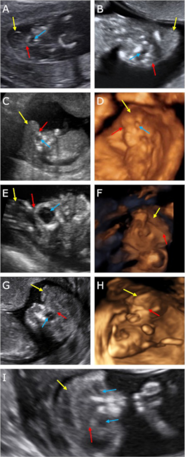
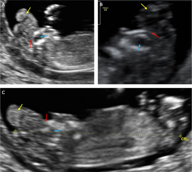
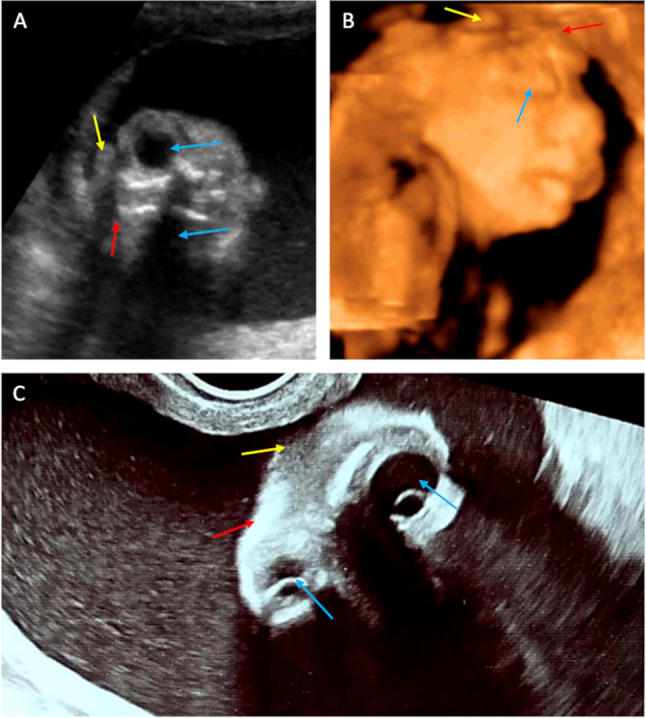
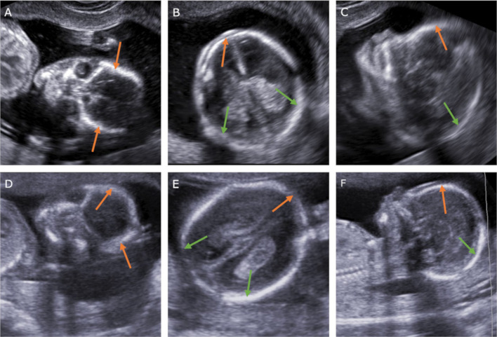
Results: In 10 cases, the absence of calvarium was diagnosed, allowing recognition of either AEAS stages or primary anencephaly. In 5 cases, cerebral structures were enclosed by an inertial rippled thin membrane, with a smooth outer contour. Between the described membrane and the brain structures, a thin anechoic space corresponding to cerebrospinal fluid was observed. This sign was defined as the "beret" sign. In these cases, acrania was diagnosed. In three cases calvarium was missing. The brain structures had an irregular appearance, did not wave and remained motionless. The outer contour was unequal as if divided into lobes. Amniotic fluid was anechoic. Exencephaly was diagnosed in these cases. In two cases calvarium, brain structures, and meninges were missing. The "frog eyes" sign and slightly echogenic amniotic fluid were visible. In this case, anencephaly was diagnosed.
Conclusions: The "beret" sign seems to be a promising tool in the diagnosis of acrania. Furthermore, echogenicity of amniotic fluid could be useful during differentiation between primary and secondary anencephaly.
Keywords: Acrania; Acrania–exencephaly–anencephaly sequence; Anencephaly; Exencephaly; Neural tube defects; Prenatal diagnosis; “Beret” sign; “Frog-eye” sign; “Mickey mouse” sign.
Conflict of interest statement
The authors have no conflicts of interest to declare.
Figures
Similar articles
-
First-trimester echogenic amniotic fluid in the acrania-anencephaly sequence.J Ultrasound Med. 2003 Oct;22(10):1075-9; quiz 1080-1. doi: 10.7863/jum.2003.22.10.1075. J Ultrasound Med. 2003. PMID: 14606564
-
Acrania-exencephaly-anencephaly sequence phenotypic characterization using two- and three-dimensional ultrasound between 11 and 13 weeks and 6 days of gestation.J Ultrason. 2018;18(74):240-246. doi: 10.15557/JoU.2018.0035. J Ultrason. 2018. PMID: 30451407 Free PMC article.
-
Exencephaly-anencephaly sequence: proof by ultrasound imaging and amniotic fluid cytology.J Matern Fetal Med. 1996 Jul-Aug;5(4):182-5. doi: 10.1002/(SICI)1520-6661(199607/08)5:4<182::AID-MFM4>3.0.CO;2-G. J Matern Fetal Med. 1996. PMID: 8796791
-
Exencephaly-anencephaly Sequence.Am J Obstet Gynecol. 2020 Dec;223(6):B5-B8. doi: 10.1016/j.ajog.2020.08.176. Epub 2020 Nov 7. Am J Obstet Gynecol. 2020. PMID: 33168213 Review. No abstract available.
-
From exencephaly to anencephaly: a catastrophic continuum of neural tube defects from embryogenesis to ultrasonographic diagnosis.J Perinat Med. 2025 Aug 5. doi: 10.1515/jpm-2025-0125. Online ahead of print. J Perinat Med. 2025. PMID: 40760857 Review.
Cited by
-
Fetal acrania diagnosed at 17 weeks of gestation by 2D∕3D ultrasound: a case report and literature review.Rom J Morphol Embryol. 2024 Jan-Mar;65(1):125-129. doi: 10.47162/RJME.65.1.16. Rom J Morphol Embryol. 2024. PMID: 38527993 Free PMC article. Review.
-
Anencephaly in a triplet pregnancy: Unprecedented spontaneous reabsorption in-utero and subsequent normal delivery via c-section: A rare case report.Radiol Case Rep. 2024 Apr 24;19(7):2826-2831. doi: 10.1016/j.radcr.2024.03.063. eCollection 2024 Jul. Radiol Case Rep. 2024. PMID: 38689815 Free PMC article.
-
Research progress on ultrasound and molecular markers for prenatal diagnosis of neural tube defects.Heliyon. 2024 Aug 13;10(16):e36060. doi: 10.1016/j.heliyon.2024.e36060. eCollection 2024 Aug 30. Heliyon. 2024. PMID: 39247260 Free PMC article. Review.
-
Prenatal Diagnosis of Acrania in One Twin of a Dichorionic Diamniotic Pregnancy: A Case Report on Management and Perinatal Outcome.Reports (MDPI). 2025 May 22;8(2):75. doi: 10.3390/reports8020075. Reports (MDPI). 2025. PMID: 40710866 Free PMC article.
References
-
- Kitova T, Milkov D, Kitov B, et al. Demographic factors and associated anomalies in fetuses with neural tube defects. Pteridines. 2013;24:257–263. doi: 10.1515/pterid-2013-0028. - DOI
-
- Karasu Y, Bozkurt M, Gençdal S et al (2015) Prenatal diagnosis of fetal acrania using two and three dimensional ultrasound. Proc Obstet Gynecol 5(1):4. 10.17077/2154-4751.1281
Publication types
MeSH terms
Substances
LinkOut - more resources
Full Text Sources
Medical
Research Materials
