

The importance of multisection sagittal and coronal magnetic resonance imaging evaluation in the assessment of temporomandibular joint disc position
- PMID: 32556656
- PMCID: PMC7785556
- DOI: 10.1007/s00784-020-03347-9
The importance of multisection sagittal and coronal magnetic resonance imaging evaluation in the assessment of temporomandibular joint disc position
Abstract
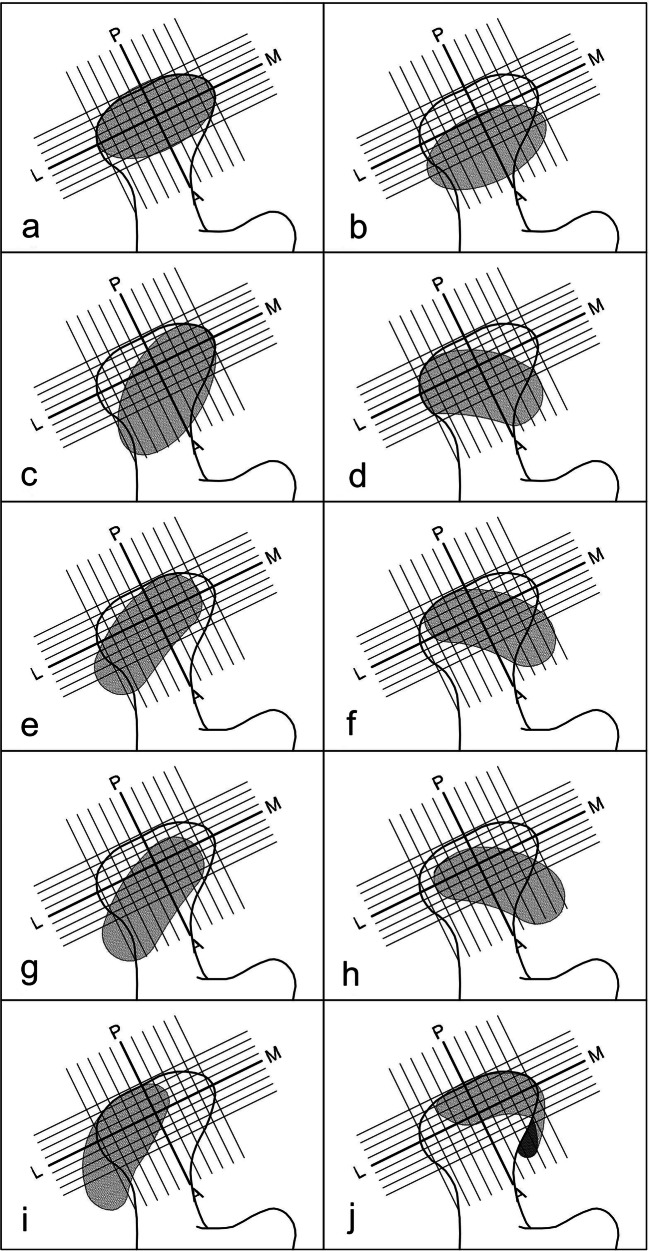
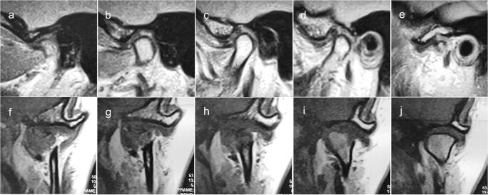
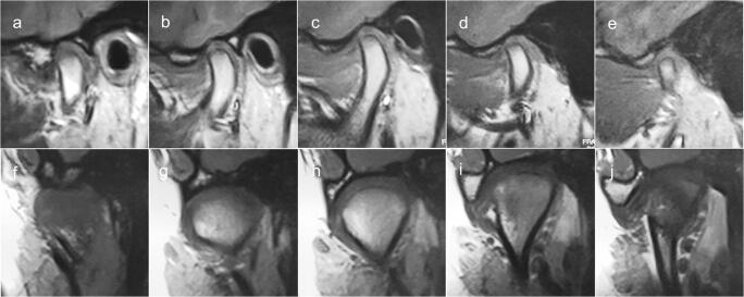
Objectives: The aim of this study was to evaluate diagnoses of temporomandibular (TMJ) disc displacement by comparing evaluations done on the basis of central sagittal scans only, the most often used in temporomandibular disorder (TMD) patients, with a multisection evaluation done with both sagittal and coronal scans.
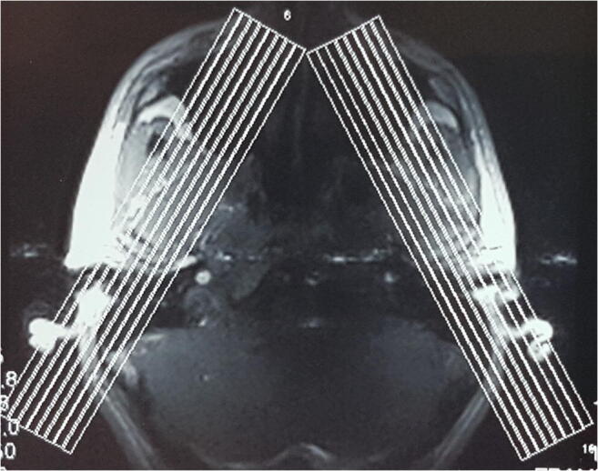
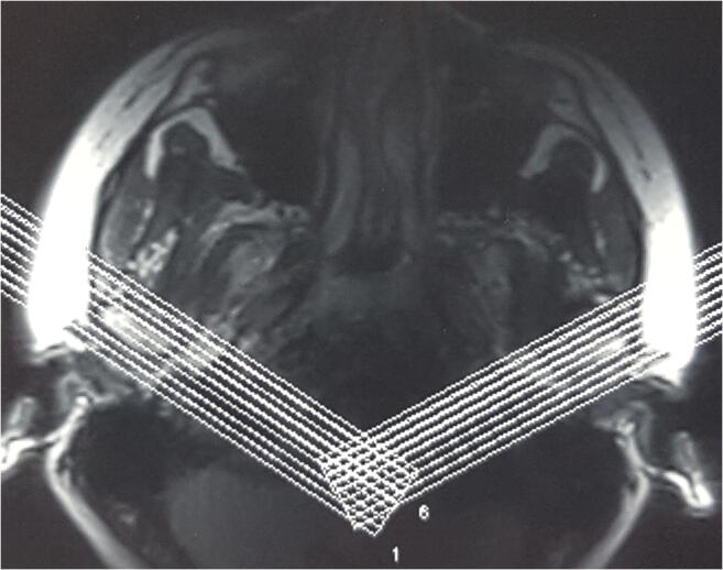
Materials and methods: Multisection MRI analysis of 382 TMJs was conducted in 191 patients with disc displacement according to RDC/TMD criteria. Disc position in the intercuspal position (IP) was assessed two times using two different methods. The first method involved a TMJ disc position evaluation on the central slide in the oblique sagittal plane only. In the second method, the TMJ disc position was assessed on all oblique sagittal and coronal images. McNemar's χ2 test was used to evaluate the differences between the sensitivities of two methods.
Results: The first method (central oblique sagittal scans assessment) identified 148 TMJs (38.7%) with normal disc position compared with 89 TMJs (23.3%) with normal disc position found by the second method (all oblique sagittal and coronal scans assessment). The sensitivity of analysis in both planes was significantly higher than in the sagittal plane only (p < 0.001).
Conclusions: The multisection analysis in the sagittal and coronal plane allows to distinguish the correct disc position from disc displacement and thus improve evaluation of TMJ internal derangement.
Clinical relevance: The multisection sagittal and coronal images should be recommended as a standard in MRI of the TMJ disc displacement in patients with TMD to avoid false-negative diagnoses.
Keywords: Magnetic resonance imaging; Temporomandibular joint disc; Temporomandibular joint disorders; Temporomandibular joint dysfunction syndrome.
Conflict of interest statement
The authors declare that they have no conflict of interest.
Figures
Similar articles
-
Correlation between direction and severity of temporomandibular joint disc displacement and reduction ability during mouth opening.J Oral Rehabil. 2017 Dec;44(12):957-963. doi: 10.1111/joor.12576. Epub 2017 Oct 6. J Oral Rehabil. 2017. PMID: 28940680
-
[Evaluation of the relationship between the attachment type of lateral pterygoid muscle and the position of temporomandibular joint disc in patients with temporomandibular joint disorders based on wireless amplified MRI detector high resolution imaging].Zhonghua Kou Qiang Yi Xue Za Zhi. 2023 Jun 9;58(6):569-574. doi: 10.3760/cma.j.cn112144-20230418-00161. Zhonghua Kou Qiang Yi Xue Za Zhi. 2023. PMID: 37272002 Chinese.
-
Disk and joint morphology variations on coronal and sagittal MRI in temporomandibular joint disorders.Clin Oral Investig. 2013 May;17(4):1243-50. doi: 10.1007/s00784-012-0803-4. Epub 2012 Aug 7. Clin Oral Investig. 2013. PMID: 22868824
-
Disk position and the bilaminar zone of the temporomandibular joint in asymptomatic young individuals by magnetic resonance imaging.Oral Surg Oral Med Oral Pathol Oral Radiol Endod. 2002 Sep;94(3):372-8. doi: 10.1067/moe.2002.127086. Oral Surg Oral Med Oral Pathol Oral Radiol Endod. 2002. PMID: 12324796 Review.
-
Role of magnetic resonance imaging in the clinical diagnosis of the temporomandibular joint.Cells Tissues Organs. 2005;180(1):6-21. doi: 10.1159/000086194. Cells Tissues Organs. 2005. PMID: 16088129 Review.
Cited by
-
Influence of examiner calibration on clinical and MRI diagnosis of temporomandibular joint disc displacement: a systematic review and meta-analysis.Dentomaxillofac Radiol. 2024 Sep 1;53(6):341-353. doi: 10.1093/dmfr/twae027. Dentomaxillofac Radiol. 2024. PMID: 38970385 Free PMC article.
-
Relationship between Clinical Symptoms and Magnetic Resonance Imaging in Temporomandibular Disorder (TMD) Patients Utilizing the Piper MRI Diagnostic System.J Clin Med. 2021 Oct 13;10(20):4698. doi: 10.3390/jcm10204698. J Clin Med. 2021. PMID: 34682820 Free PMC article.
-
Magnetic Resonance Imaging Evaluation of Closed-Mouth TMJ Disc-Condyle Relationship in a Population of Patients Seeking for Temporomandibular Disorders Advice.Pain Res Manag. 2021 Dec 2;2021:5565747. doi: 10.1155/2021/5565747. eCollection 2021. Pain Res Manag. 2021. PMID: 34900071 Free PMC article.
-
COL12A1 Single Nucleotide Polymorphisms rs240736 and rs970547 Are Not Associated with Temporomandibular Joint Disc Displacement without Reduction.Genes (Basel). 2021 May 5;12(5):690. doi: 10.3390/genes12050690. Genes (Basel). 2021. PMID: 34062975 Free PMC article.
-
Comparison of magnetic resonance imaging findings in 880 temporomandibular disorder patients of different age groups: a retrospective study.BMC Oral Health. 2022 Dec 28;22(1):651. doi: 10.1186/s12903-022-02666-5. BMC Oral Health. 2022. PMID: 36577982 Free PMC article.
References
-
- Schiffman E, Ohrbach R, Truelove E, Look J, Anderson G, Goulet JP, List T, Svensson P, Gonzalez Y, Lobbezoo F, Michelotti A, Brooks SL, Ceusters W, Drangsholt M, Ettlin D, Gaul C, Goldberg LJ, Haythornthwaite JA, Hollender L, Jensen R, John MT, De Laat A, de Leeuw R, Maixner W, van der Meulen M, Murray GM, Nixdorf DR, Palla S, Petersson A, Pionchon P, Smith B, Visscher CM, Zakrzewska J, Dworkin SF, InternationalRDC/TMDConsortiumNetwork, International Association for Dental Research; Orofacial PainSpecialInterest Group, International Association for the Study of Pain Diagnostic Criteria for Temporomandibular Disorders (DC/TMD) for clinical and research applications: recommendations of the International RDC/TMD Consortium Network and Orofacial Pain Special Interest Group. J Oral Facial Pain Headache. 2014;28:6–27. doi: 10.11607/jop.1151. - DOI - PMC - PubMed
-
- Manfredini D, Guarda-Nardini L. Agreement between Research Diagnostic Criteria for Temporomandibular Disorders and magnetic resonance diagnoses of temporomandibular disc displacement in a patient population. Int J Oral Maxillofac Surg. 2008;37:612–616. doi: 10.1016/j.ijom.2008.04.003. - DOI - PubMed
MeSH terms
LinkOut - more resources
Full Text Sources
Medical
