

Glial cell line-derived neurotrophic factors (GFLs) and small molecules targeting RET receptor for the treatment of pain and Parkinson's disease
- PMID: 32556722
- PMCID: PMC7529621
- DOI: 10.1007/s00441-020-03227-4
Glial cell line-derived neurotrophic factors (GFLs) and small molecules targeting RET receptor for the treatment of pain and Parkinson's disease
Abstract
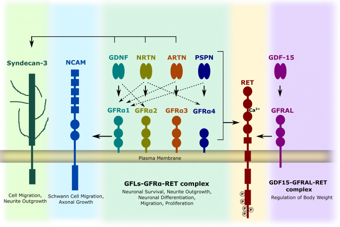
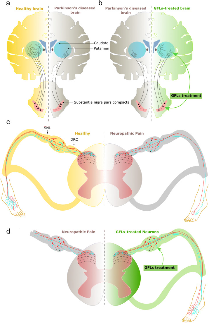
Rearranged during transfection (RET), in complex with glial cell line-derived (GDNF) family receptor alpha (GFRα), is the canonical signaling receptor for GDNF family ligands (GFLs) expressed in both central and peripheral parts of the nervous system and also in non-neuronal tissues. RET-dependent signaling elicited by GFLs has an important role in the development, maintenance and survival of dopamine and sensory neurons. Both Parkinson's disease and neuropathic pain are devastating disorders without an available cure, and at the moment are only treated symptomatically. GFLs have been studied extensively in animal models of Parkinson's disease and neuropathic pain with remarkable outcomes. However, clinical trials with recombinant or viral vector-encoded GFL proteins have produced inconclusive results. GFL proteins are not drug-like; they have poor pharmacokinetic properties and activate multiple receptors. Targeting RET and/or GFRα with small molecules may resolve the problems associated with using GFLs as drugs and can result in the development of therapeutics for disease-modifying treatments against Parkinson's disease and neuropathic pain.
Keywords: Drug design; Drug development; GDNF family ligands (GFLs); Glial cell line-neurotrophic factor (GDNF); Neurturin (NRTN); RET agonist; RET receptor tyrosine kinase; RET receptor, artemin (ARTN); Small molecule.
Conflict of interest statement
Dr. Sidorova is a minor shareholder in GeneCode Ltd., a company owning IPRs for RET agonists from the BT13 family. The other author declares that he has no competing interests.
Figures
References
-
- Airaksinen MS, Saarma M. The GDNF family: signalling, biological functions and therapeutic value. Nat Rev Neurosci. 2002;3:383–394. - PubMed
-
- Åkerud P, Holm PC, Castelo-Branco G, et al. Persephin-overexpressing neural stem cells regulate the function of nigral dopaminergic neurons and prevent their degeneration in a model of Parkinson’s disease. Mol Cell Neurosci. 2002;21:205–222. - PubMed
Publication types
MeSH terms
Substances
Grants and funding
LinkOut - more resources
Full Text Sources
Medical
