

Optical Flow Methods for Lung Nodule Segmentation on LIDC-IDRI Images
- PMID: 32556911
- PMCID: PMC7572960
- DOI: 10.1007/s10278-020-00346-w
Optical Flow Methods for Lung Nodule Segmentation on LIDC-IDRI Images
Abstract
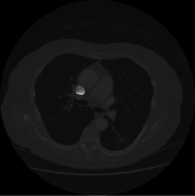
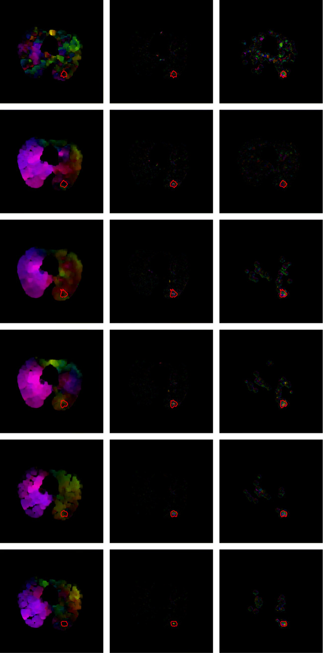
Lung nodule segmentation is an essential step in any CAD system for lung cancer detection and diagnosis. Traditional approaches for image segmentation are mainly morphology based or intensity based. Motion-based segmentation techniques tend to use the temporal information along with the morphology and intensity information to perform segmentation of regions of interest in videos. CT scans comprise of a sequence of dicom 2-D image slices similar to videos which also comprise of a sequence of image frames ordered on a timeline. In this work, Farneback, Horn-Schunck and Lucas-Kanade optical flow methods have been used for processing the dicom slices. The novelty of this work lies in the usage of optical flow methods, generally used in motion-based segmentation tasks, for the segmentation of nodules from CT images. Since thin-sliced CT scans are the imaging modality considered, they closely approximate the motion videos and are the primary motivation for using optical flow for lung nodule segmentation. This paper also provides a detailed comparative analysis and validates the effectiveness of using optical flow methods for segmentation. Finally, we propose methods to further improve the efficiency of segmentation using optical flow methods on CT scans.
Keywords: Computed tomography; Optical flow; Pulmonary nodule; Segmentation.
Conflict of interest statement
The authors declare that they have no conflict of interest.
Figures


References
-
- Adams R, Bischof L. Seeded region growing. IEEE Trans Pattern Anal Mach Intell. 1994;16(6):641–647.
-
- Armato SG, III, McLennan G, Bidaut L, McNitt-Gray MF, Meyer CR, Reeves AP, Zhao B, Aberle DR, Henschke CI, Hoffman EA, et al. The lung image database consortium (lidc) and image database resource initiative (idri): a completed reference database of lung nodules on ct scans. Med Phys. 2011;38(2):915–931. - PMC - PubMed
-
- Badura P, Piętka E: Pre-and postprocessing stages in fuzzy connectedness-based lung nodule cad.. In: Information technologies in biomedicine. Springer, 2008, pp 192–199
-
- Badura P, Pietka E. Soft computing approach to 3d lung nodule segmentation in ct. Comput Biol Med. 2014;53:230–243. - PubMed
-
- Bagci U, Chen X, Udupa JK. Hierarchical scale-based multiobject recognition of 3-d anatomical structures. IEEE Trans Med Imaging. 2011;31(3):777–789. - PubMed
Publication types
MeSH terms
LinkOut - more resources
Full Text Sources
Medical
Miscellaneous
