Review
. 2020 Sep;18(9):2103-2109.
doi: 10.1111/jth.14975. Epub 2020 Jul 21.

Coagulopathy in COVID-19

Affiliations
Review

Coagulopathy in COVID-19

Toshiaki Iba et al. J Thromb Haemost. 2020 Sep.

Abstract

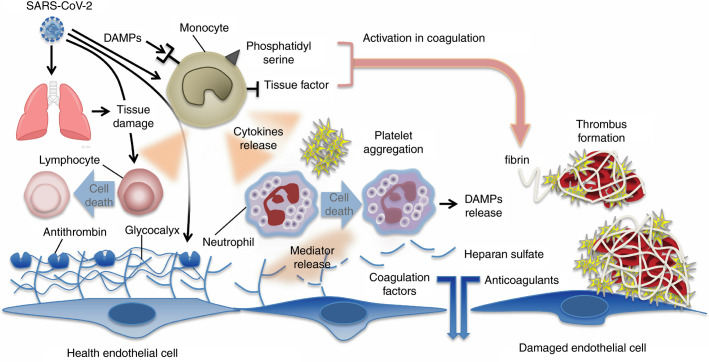
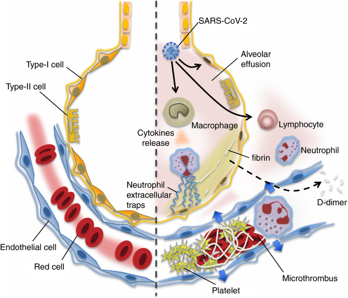
The COVID-19 pandemic has become an urgent issue in every country. Based on recent reports, the most severely ill patients present with coagulopathy, and disseminated intravascular coagulation (DIC)-like massive intravascular clot formation is frequently seen in this cohort. Therefore, coagulation tests may be considered useful to discriminate severe cases of COVID-19. The clinical presentation of COVID-19-associated coagulopathy is organ dysfunction primarily, whereas hemorrhagic events are less frequent. Changes in hemostatic biomarkers represented by increase in D-dimer and fibrin/fibrinogen degradation products indicate the essence of coagulopathy is massive fibrin formation. In comparison with bacterial-sepsis-associated coagulopathy/DIC, prolongation of prothrombin time, and activated partial thromboplastin time, and decrease in antithrombin activity is less frequent and thrombocytopenia is relatively uncommon in COVID-19. The mechanisms of the coagulopathy are not fully elucidated, however. It is speculated that the dysregulated immune responses orchestrated by inflammatory cytokines, lymphocyte cell death, hypoxia, and endothelial damage are involved. Bleeding tendency is uncommon, but the incidence of thrombosis in COVID-19 and the adequacy of current recommendations regarding standard venous thromboembolic dosing are uncertain.

Keywords: COVID-19; anticoagulant; coagulopathy; coronavirus; disseminated intravascular coagulation.


Figures

Figure 1
Figure 1
Mechanisms of coagulation activation in Covid‐19. Both pathogens (viruses) and damage‐associated molecular patterns (DAMPs) from injured host tissue can activate monocytes. Activated monocytes release inflammatory cytokines and chemokines that stimulate neutrophils, lymphocytes, platelets, and vascular endothelial cells. Monocytes and other cells express tissue factor and phosphatidylserine on their surfaces and initiate coagulation. Healthy endothelial cells maintain their anti‐thrombogenicity by expressing glycocalyx and its binding protein antithrombin. Damaged endothelial cells change their properties to procoagulant following disruption of the glycocalyx and loss of anticoagulant proteins
Figure 2
Figure 2
In the undamaged lung (left), smooth blood flow and the effective oxygenation is recognized. Covid‐19 infection causes an intense inflammatory reaction (right). The lung tissue damages are induced by uncontrolled activation of lymphocytes and possibly neutrophil activation (neutrophil extracellular traps formation). Increased pulmonary production of platelets is also involved in the defense process. In the damaged lung, the virulence of Covid‐19 or unabated inflammatory reaction causes pulmonary microthrombi, endothelial damage, and vascular leakage. The host intends to control the thrombi formation by vigorous fibrinolysis because lung has high fibrinolytic capacity. The fibrin degraded fragment (D‐dimer) spills into the blood and is detected in the blood samples

Comment in

  • Comment on an article: "Coagulopathy in COVID-19".
    Ibrahimagić OĆ, Smajlović D, Dostović Z, Kunić S, Šehanović A, Kojić B. Ibrahimagić OĆ, et al. J Thromb Haemost. 2020 Dec;18(12):3381-3382. doi: 10.1111/jth.15122. J Thromb Haemost. 2020. PMID: 33090645 Free PMC article. No abstract available.

Similar articles

Cited by

References

    1. Wu C., Chen X., Cai Y., et al. Risk factors associated with acute respiratory distress syndrome and death in patients with coronavirus disease 2019 pneumonia in Wuhan, China. JAMA Intern Med. 2019;2020 - PMC - PubMed
    1. Schnittler H.J., Feldmann H. Viral hemorrhagic fever–a vascular disease? Thromb Haemost. 2003;89(6):967–972. - PubMed
    1. Paessler S., Walker D.H. Pathogenesis of the viral hemorrhagic fevers. Annu Rev Pathol. 2013;24(8):411–440. - PubMed
    1. Falasca L., Agrati C., Petrosillo N., et al. Molecular mechanisms of Ebola virus pathogenesis: focus on cell death. Cell Death Differ. 2015;22(8):1250–1259. - PMC - PubMed
    1. Wong R.S., Wu A., To K.F., et al. Haematological manifestations in patients with severe acute respiratory syndrome: retrospective analysis. BMJ. 2003;326:1358–1362. - PMC - PubMed

Publication types

MeSH terms