. 2020 Oct;40(10):2455-2468.
doi: 10.1111/liv.14569. Epub 2020 Jul 7.

Senescent cholangiocytes release extracellular vesicles that alter target cell phenotype via the epidermal growth factor receptor

Affiliations

Senescent cholangiocytes release extracellular vesicles that alter target cell phenotype via the epidermal growth factor receptor

Mohammed S Al Suraih et al. Liver Int. 2020 Oct.

Abstract

Background & aims: Primary sclerosing cholangitis (PSC) is a chronic liver disease characterized by peribiliary inflammation and fibrosis. Cholangiocyte senescence is a prominent feature of PSC. Here, we hypothesize that extracellular vesicles (EVs) from senescent cholangiocytes influence the phenotype of target cells.

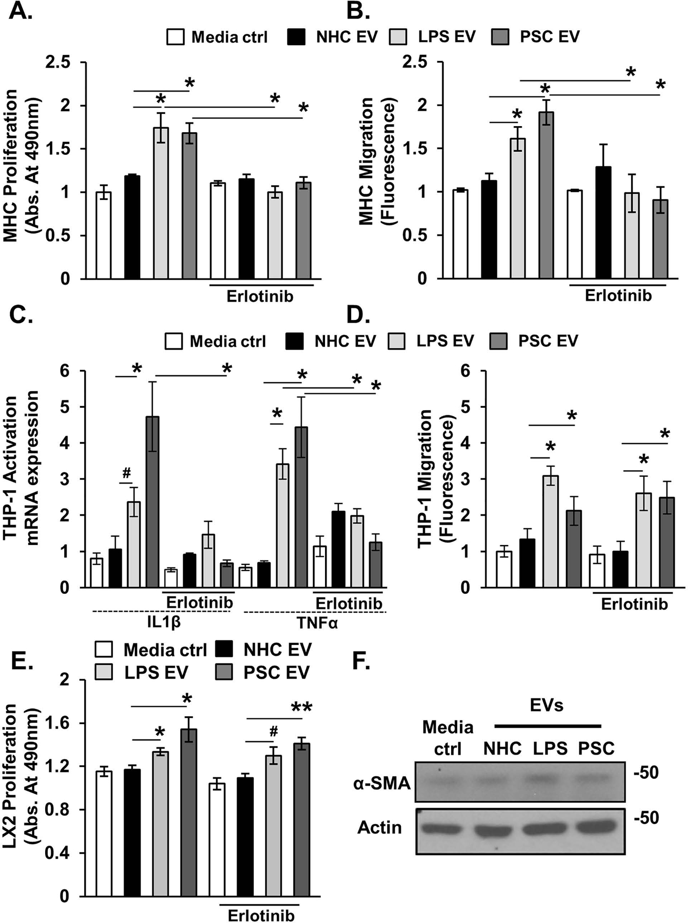
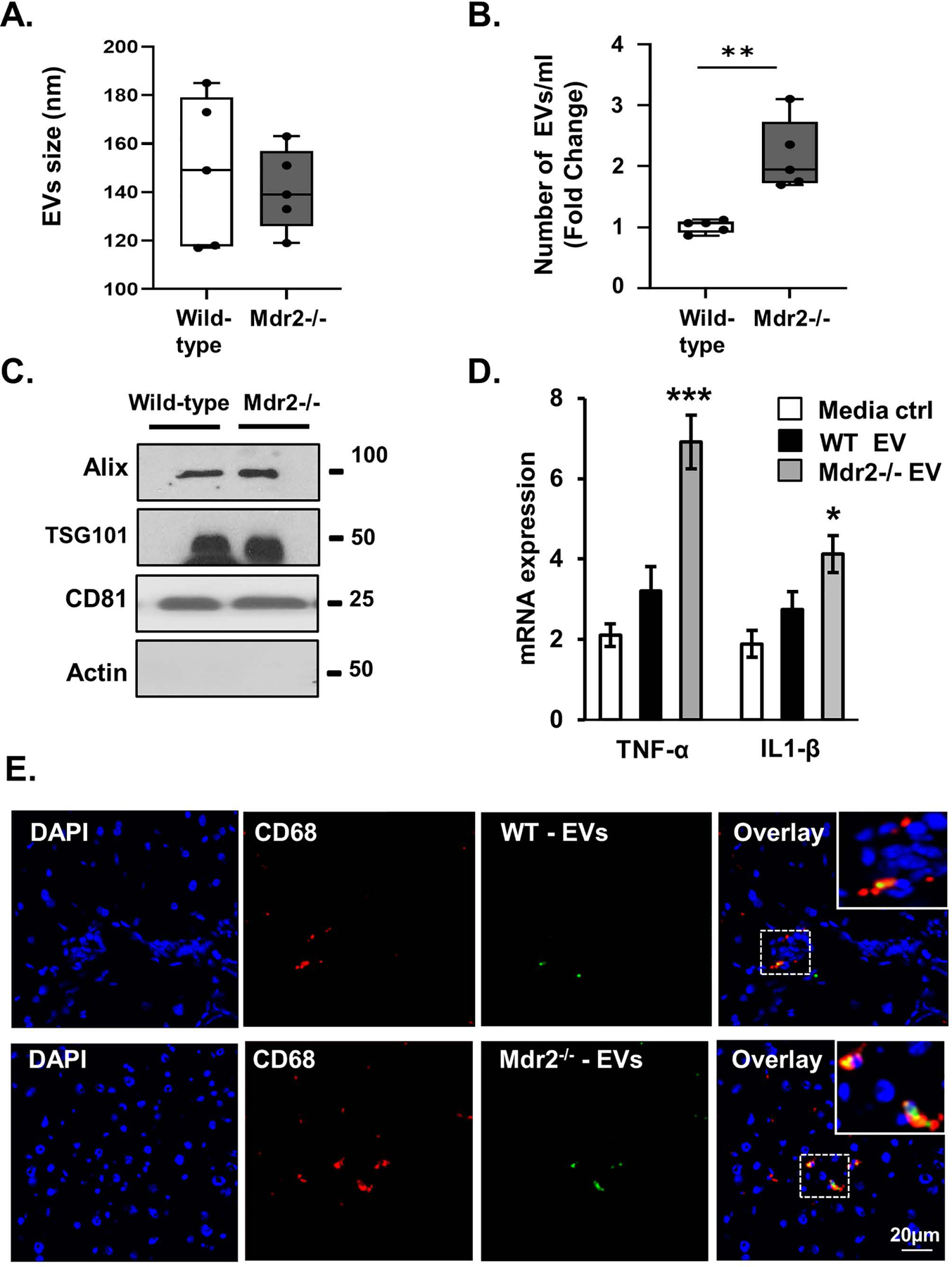
Methods: EVs were isolated from normal human cholangiocytes (NHCs), cholangiocytes from PSC patients and NHCs experimentally induced to senescence. NHCs, malignant human cholangiocytes (MHCs) and monocytes were exposed to 108 EVs from each donor cell population and assessed for proliferation, MAPK activation and migration. Additionally, we isolated EVs from plasma of wild-type and Mdr2-/- mice (a murine model of PSC), and assessed mouse monocyte activation.

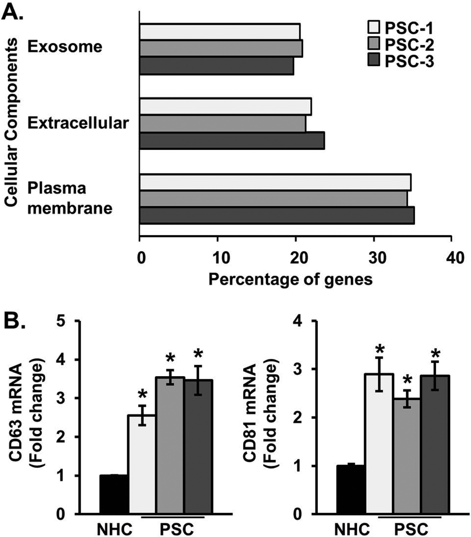
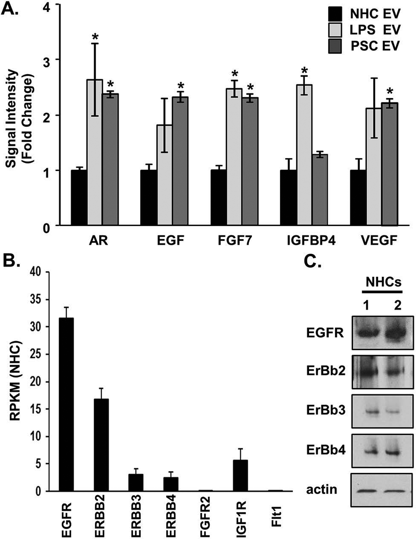
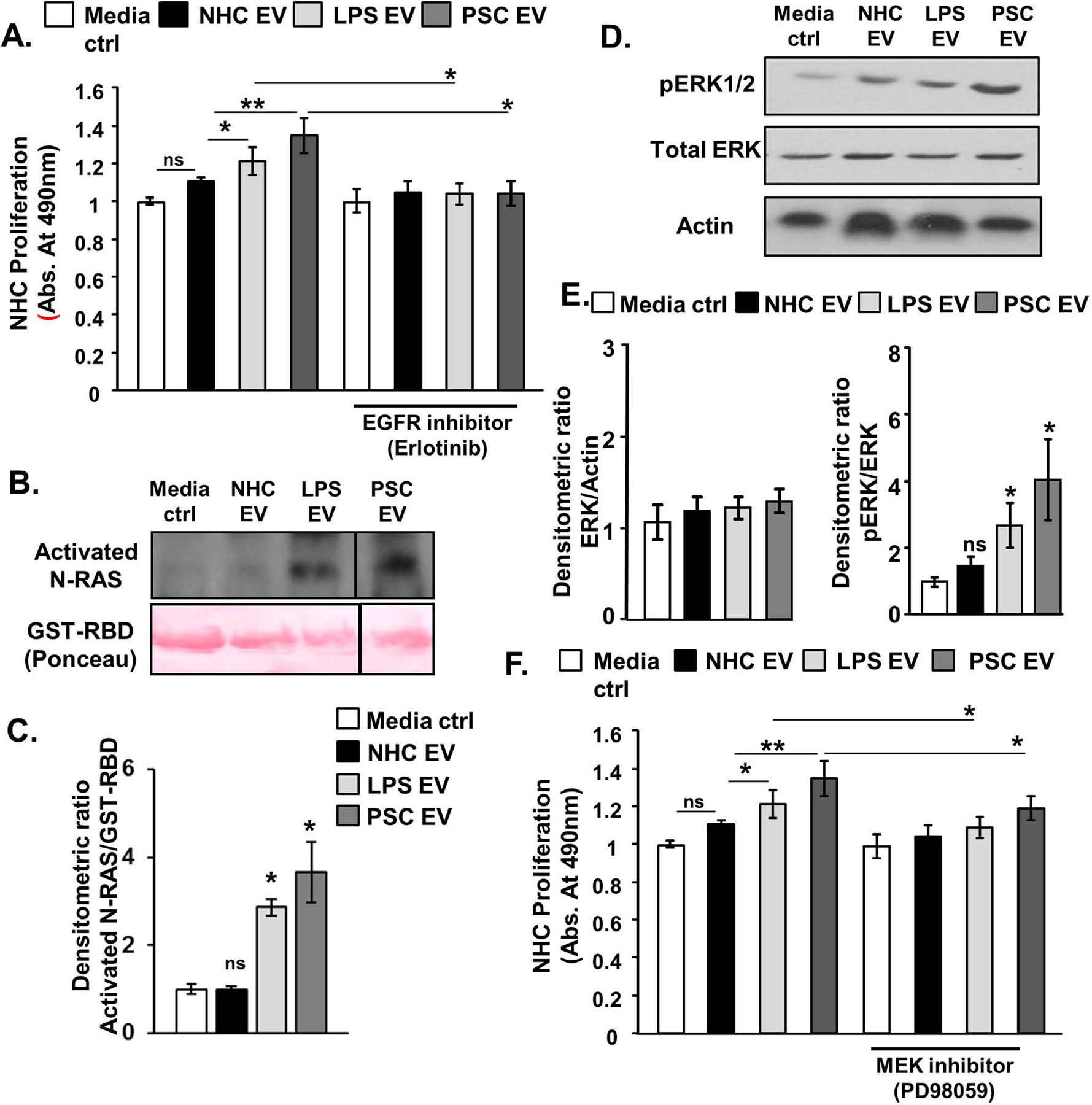
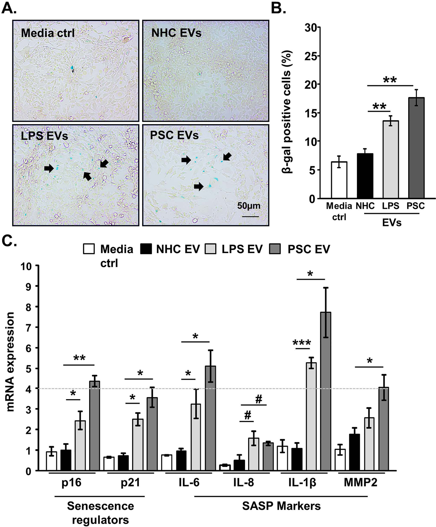
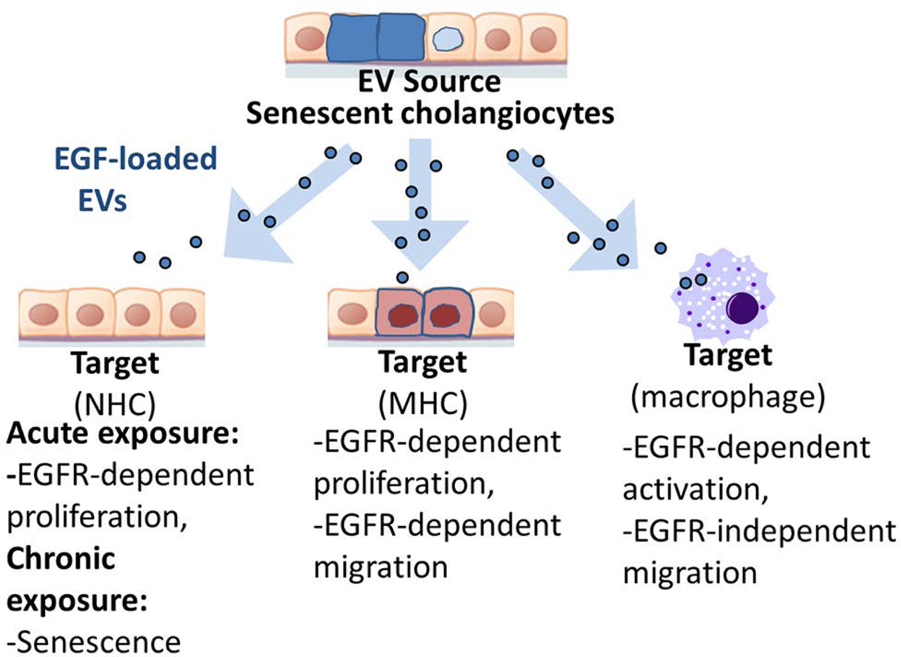
Results: EVs exhibited the size and protein markers of exosomes. The number of EVs released from senescent human cholangiocytes was increased; similarly, the EVs in plasma from Mdr2-/- mice were increased. Additionally, EVs from senescent cholangiocytes were enriched in multiple growth factors, including EGF. NHCs exposed to EVs from senescent cholangiocytes showed increased NRAS and ERK1/2 activation. Moreover, EVs from senescent cholangiocytes promoted proliferation of NHCs and MHCs, findings that were blocked by erlotinib, an EGF receptor inhibitor. Furthermore, EVs from senescent cholangiocytes induced EGF-dependent Interleukin 1-beta and Tumour necrosis factor expression and migration of human monocytes; similarly, Mdr2-/- mouse plasma EVs induced activation of mouse monocytes.

Conclusions: The data continue to support the importance of cholangiocyte senescence in PSC pathogenesis, directly implicate EVs in cholangiocyte proliferation, malignant progression and immune cell activation and migration, and identify novel therapeutic approaches for PSC.

Keywords: biliary epithelial cell; cellular senescence; extracellular vesicles; primary sclerosing cholangitis; senescence-associated secretory phenotype.


Conflict of interest statement

Conflict of interest: The authors have no conflict of interest related to the manuscript.

Figures

Figure 1.
Figure 1.. Primary sclerosing cholangitis (PSC) patient-derived cholangiocytes are enriched in exosomes.
A, RNA sequencing was performed on one normal human cholangiocyte (NHC) and three PSC patient-derived short-term cholangiocyte cultures. Genes expressed at a level of at least 5 copies per million in NHC, and upregulated more than 2-fold in PSC cholangiocytes were used for functional enrichment analysis. Based on these criteria, the cholangiocytes isolated from the three PSC patients (PSC-1, PSC-2, and PSC-3) exhibited increased expression of 866, 1,026, and 877 genes compared to NHC, respectively. We identified consistent enrichment in the cellular components categories of exosome, extracellular, and plasma membrane. Bars show percentage, ie, number of upregulated genes in each category/total number of upregulated genes. The hypergeometric P value test was performed for each PSC patient-derived sample in each category and ranged from 9.5E-19 - 2.0E-05. B, Messenger RNA (mRNA) expression of exosomal markers (CD63 and CD81) are elevated in PSC patient-derived cholangiocytes. Bars represent mean fold-change of per million mapped reads (RPKM) vs NHC (±) standard error of the mean (SEM); n=3. *P<0.05 vs NHC.
Figure 2.
Figure 2.. Cultured senescent cholangiocytes secrete an increased number of extracellular vesicles (EVs).
A, EVs isolated from normal and senescent cholangiocytes (lipopolysaccharide- [LPS-] induced, and primary sclerosing cholangitis [PSC]-patient derived) were examined by transmission electron microscopy (EM) and immunogold electron microscopy (IEM). The purified vesicles exhibited the typical “deflated football” exosome morphology (inset) and were positive for the exosomal marker, CD63. B, Immunoblotting on whole cell (WCL) and isolated EV lysates. Analysis indicated that EV samples are positive for exosomal markers, Alix, TSG101, and CD9, and are negative for the cytoskeletal protein, actin. C, Nanoparticle tracking analysis (NTA) of NHC (dotted line), LPS-induced (dashed line) and PSC patient-derived senescent cholangiocytes (solid line) D, and E, Quantitation of NTA showed similar EV size between NHCs and LPS-, H202-, irradiation (IR)-induced or PSC patient-derived senescent cholangiocytes. However, the number of EVs, normalized to total cell count, was increased in the senescent cholangiocytes. Bars represent mean (±) standard error of the mean (SEM); n=7–10. *P<0.001.
Figure 3.
Figure 3.. Senescent cholangiocyte-derived extracellular vesicles (EVs) are enriched in growth factors.
A, Equal numbers of normal and senescent cholangiocyte-derived EVs were harvested for protein analysis using a growth factor array (Abcam). Bars represent mean (±) standard error of the mean (SEM); n=3.The primary sclerosing cholangitis (PSC) values represent the mean of 3 PSC patient samples. Values represent a fold change versus EVs derived from normal human cholangiocytes (NHCs). *P<0.05. B, RNA-sequencing data on NHCs, shown as reads per Kilobase of transcript, per million mapped reads (RPKM) from 3 biological replicates revealed increased expression of the epidermal growth factor receptors (EGFRs), EGFR and Erb-B2 receptor tyrosine kinase 2 (ERBB2), compared to the fibroblast growth factor receptor (FGFR2), the insulin-like growth factor 1 receptor (IGF1R), and FMS-related tyrosine kinase 1 (FLT1, ie, VEGFR-1). C, Immunoblotting analysis on NHCs (2 biological replicates) showed positive protein expression of the EGF receptors.
Figure 4.
Figure 4.. Senescent cholangiocyte-derived extracellular vesicles (EVs) induce proliferation of bystander cholangiocytes.
A, MTS proliferation assay; equal number of normal and senescent cholangiocytes derived EVs were applied to normal human cholangiocytes (NHCs) in the presence of absence of the epidermal growth factor receptor inhibitor, erlotinib. Cellular proliferation was measured after 24 hours incubation. The observed increase in proliferation in NHCs treated with senescent derived EVs is abrogated in the presence of erlotinib. B and C, NRAS activation assay and semi-quantitative densitometric analysis demonstrated that EVs isolated from senescent cholangiocytes (1e8/ml) promoted NRAS activation. D and E, Immunoblotting analysis and semi-quantitative densitometric analysis indicated that senescent cholangiocytes derived EVs (1e8/ml) induced increased ERK1/2 phosphorylation. F, MTS proliferation assay; equal number of normal and senescent cholangiocytes derived EVs were applied to NHCs in the presence of absence of the (MEK inhibitor, PD98059. Cellular proliferation was measured after 24 hours incubation. The observed increase in proliferation in NHCs treated with senescent derived EVs is abrogated in the presence of PD98059. For all panels, bars represent mean (±) standard error of the mean (SEM); n=3–5 biological replicates, *P<0.05, **P<0.01.
Figure 5.
Figure 5.. Senescent cholangiocyte-derived extracellular vesicles (EVs) induce cellular senescence of bystander cholangiocytes.
A, Senescence-associated β-galactosidase activity (SA-β-gal) assay; bystander normal human cholangiocytes (NHCs) were incubated with normal and senescent (lipopolysaccharide [LPS]-induced, and primary sclerosing cholangitis [PSC]-derived) cholangiocyte-derived EVs (1e8/ml) every other day for 10 days. SA-β-gal activity was assessed using β-galactosidase assay. Arrows indicate representative SA-β-gal positive NHCs. B, Quantification of the SA-β-gal assay; data presented as percentage of SA-β-gal positive cells per total cell count. The PSC patient-derived data represent the average of 3 replicates of 3–5 separate PSC patient samples. C, Induction of senescence was assessed by measuring messenger RNA (mRNA) expression (RT-qPCR) of various cellular senescence markers. Bars represent mean (±) standard error of the mean (SEM); n=3–5. *P<0.05, **P<0.01, #P=0.07.
Figure 6.
Figure 6.. Senescent cholangiocyte-derived extracellular vesicles (EVs) promote phenotypic changes in target cells.
Equal number of normal and senescent cholangiocyte-derived EVs were applied to MHCs (A&B, Hucct1), human monocytes (C&D, THP-1), and hepatic stellate cells (E&F, LX2) for 24 hours incubation period (+/− erlotinib). A, MTS proliferation assay. EVs isolated from senescent cholangiocytes induced an increase in the target MHCs proliferation. Incubation with erlotinib prevented senescent EV-induced increased proliferation. B, Migration was measured using a colorimetric assay (Cell Biolabs). Senescent-derived EVs promoted MHC migration, while erlotinib inhibited this effect. C, Activation of THP-1 human monocytes was assessed by measuring monocytes messenger RNA (mRNA) expression levels of IL1B and TNF. Senescent-derived EVs promoted expression of these proinflammatory genes, while erlotinib treatment blunted this response. D, Migration of THP-1. Senescent-derived EVs promoted THP-1 migration and erlotinib had no effect on senescent EV-induced THP-1 migration. E, MTS proliferation assay. Senescent cholangiocyte-derived EVs induced proliferation of LX2. F, Immunoblotting analysis of α-SMA (stellate cells activation marker) showed no difference in expression upon incubation with senescent-derived EVs. Bars represent mean (±) standard error of the mean (SEM); n=3. *P<0.05, **P<0.01, #p=0.07.
Figure 7.
Figure 7.. Circulating extracellular vesicles (EVs) are increased in the Mdr2−/− murine model of primary sclerosing cholangitis (PSC).
A and B, Characterization of wild-type C57BL/6J and Mdr2−/− mouse plasma-derived EVs, vesicle size, and number using nanoparticle tracking analysis (NTA) S300. The EVs isolated from wild-type and Mdr2−/− were similar size, yet more EVs were detected in the plasma of Mdr2−/− mice compared to wild-type mice. Data represent size and number from 5 mice per group. C, Western blotting analysis indicated that wild-type and Mdr2−/− mouse-derived EVs were positive for the exosome marker Alix, TSG101, and CD81. D, Equal numbers of wild-type and Mdr2−/− mouse-derived EVs (1e8/ml) were applied to a mouse monocyte cell line (RAW). Activation after 24-hours incubation was assessed by measuring monocyte messenger RNA (mRNA) expression levels (RT-qPCR) of tumor necrosis factor (Tnf) and interleukin 1 beta (Il1b). E, In vivo EV uptake assay reveals that Dio dye-labeled plasma-derived EVs colocalize with hepatic monocytes. EVs were collected from wild-type (WT) or Mdr2−/− mouse plasma and labeled with DiO dye. Immunofluorescence reveals colocalization of CD68 (red, monocytes marker) and plasma EVs (green, labeled with DiO dye) in liver tissue of WT (upper panel) and Mdr2−/− (lower panel) mice. Bars represent mean (±) standard error of the mean (SEM); n=3. *P<0.05, **P<0.01.
Figure 8.
Figure 8.. Working model of intercellular communication between senescent cholangiocytes and target cells.
Senescent cholangiocytes release increased numbers of EVs that are likely enriched in exosomes. Compared to EVs from nonsenescent cholangiocytes, EVs from patient-derived or experimentally induced senescent cholangiocytes are increased in number, enriched in growth factors including epidermal growth factor receptor (EGFR) ligands, and have different functional effects on target cells.

References

    1. Lazaridis KN, LaRusso NF. Primary Sclerosing Cholangitis. N Engl J Med 2016; 375(12): 1161–70. - PMC - PubMed
    1. Bashir MR, Muir AJ. Great Expectations - Can Magnetic Resonance Elastography Accelerate Progress in Primary Sclerosing Cholangitis Research? Clin Gastroenterol Hepatol 2019. - PubMed
    1. O’Hara SP, Tabibian JH, Splinter PL, LaRusso NF. The dynamic biliary epithelia: molecules, pathways, and disease. J Hepatol 2013; 58(3): 575–82. - PMC - PubMed
    1. O’Hara SP, Karlsen TH, LaRusso NF. Cholangiocytes and the environment in primary sclerosing cholangitis: where is the link? Gut 2017; 66(11): 1873–77. - PMC - PubMed
    1. Guicciardi ME, Trussoni CE, LaRusso NF, Gores GJ. The Spectrum of Reactive Cholangiocytes in Primary Sclerosing Cholangitis. Hepatology 2019. - PMC - PubMed

Publication types