. 2020 Sep;26(9):2054-2063.
doi: 10.3201/eid2609.201495. Epub 2020 Jun 19.

Isolation, Sequence, Infectivity, and Replication Kinetics of Severe Acute Respiratory Syndrome Coronavirus 2

Isolation, Sequence, Infectivity, and Replication Kinetics of Severe Acute Respiratory Syndrome Coronavirus 2

Arinjay Banerjee et al. Emerg Infect Dis. 2020 Sep.

Abstract

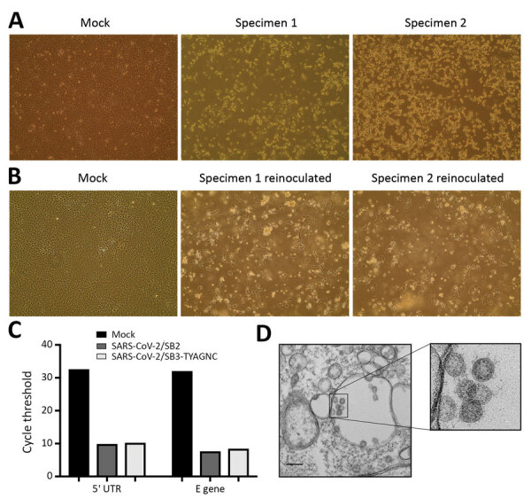
Since its emergence in Wuhan, China, in December 2019, severe acute respiratory syndrome coronavirus 2 (SARS-CoV-2) has infected ≈6 million persons worldwide. As SARS-CoV-2 spreads across the planet, we explored the range of human cells that can be infected by this virus. We isolated SARS-CoV-2 from 2 infected patients in Toronto, Canada; determined the genomic sequences; and identified single-nucleotide changes in representative populations of our virus stocks. We also tested a wide range of human immune cells for productive infection with SARS-CoV-2. We confirm that human primary peripheral blood mononuclear cells are not permissive for SARS-CoV-2. As SARS-CoV-2 continues to spread globally, it is essential to monitor single-nucleotide polymorphisms in the virus and to continue to isolate circulating viruses to determine viral genotype and phenotype by using in vitro and in vivo infection models.

Keywords: 2019 novel coronavirus disease; COVID-19; SARS-CoV-2; coronavirus disease; immune cells; isolation; phylogenetics; replication; respiratory infections; severe acute respiratory syndrome coronavirus 2; viruses; zoonoses.


Figures

Figure 1
Figure 1
Severe acute respiratory syndrome coronavirus 2 (SARS-CoV-2) protein detection in infected Vero E6 and CD4+ T cells. To detect SARS-CoV-2 protein expression, we infected Vero E6 and CD4+ T cells with SARS-CoV-2 at a multiplicity of infection of 0.1 for 24 h. We immunostained these cells and observed them by using fluorescent microscopy. A) SARS-CoV-2–infected and immunostained Vero E6 cells. B) SARS-CoV-2–infected and immunostained CD4+ T cells. For panels A and B, cells were stained by using an antibody cocktail consisting of SARS-CoV-2 S1 antibody, SARS-CoV-2 N antibody, and diluted serum from a recovered coronavirus disease patient. C) SARS-CoV-2 infected CD4+ T cells immunostained with SARS-CoV-2 S1 antibody (anti-S). Scale bars indicate 400 μm; original magnification ×10.
Figure 2
Figure 2
Isolating severe acute respiratory syndrome coronavirus 2 (SARS-CoV-2) from patients with coronavirus disease (COVID-19). A) Vero E6 cells were mock inoculated or inoculated with midturbinate clinical specimens from COVID-19 patients. Cells were incubated for 72 h and observed for cytopathic effect (CPE) under a light microscope. Original magnification ×10. B) To determine if supernatant from Vero E6 cells that were mock inoculated or inoculated with clinical specimens contained replication competent virus, we reinoculated a fresh monolayer of Vero E6 cells and observed cells under a light microscope for CPE after 24 h. Original magnification ×10. C) Quantitative real-time PCR was used to detect SARS-CoV-2 5′-UTR and E gene in RNA extracted from supernatant that was collected from Vero E6 cells that were mock infected or infected with clinical specimens from COVID-19 patients for 72 h. D) Electron micrograph of Vero E6 cells that were reinfected for 48 h with supernatant that was collected from Vero E6 cells infected with clinical specimens. Original magnification ×36,000. Inset, zoomed and cropped from the original electron micrograph, shows coronavirus-like particles. M, mock specimen; specimen 1, SARS-CoV-2/SB2; specimen 2, SARS-CoV-2/SB3-TYAGNC. E, envelope; UTR, untranslated region.
Figure 3
Figure 3
Replication of severe acute respiratory syndrome coronavirus 2 (SARS-CoV-2) in human structural and immune cells. To identify human cells that support SARS-CoV-2 replication, we infected human cell lines and primary cells at a multiplicity of infection of 0.01 (n = 2 independent experiments; supernatant from each experiment was titrated in triplicate). We infected Vero E6 cells as a control. THF (human telomerase life-extended cells) and Calu-3 cells (human lung adenocarcinoma–derived) cells represent human structural cells. THP-1 is a monocyte cell line that was used to derive macrophages and dendritic cells. PBMCs from 2 healthy human donors were used to generate CD4+, CD8+, CD19+, monocytes, and other (CD4–, CD8–, CD19–) cell populations. Supernatant from infected cells was collected at various times and titrated on Vero E6 cells to determine virus titers (TCID50). PBMC, peripheral blood mononuclear cell; TCID50, 50% tissue culture infectious dose.
Figure 4
Figure 4
Electron micrographs of severe acute respiratory syndrome coronavirus 2 (SARS-CoV-2)–infected cells. To detect coronavirus-like particles in experimentally infected human structural and immune cells, we infected a range of cells with SARS-CoV-2 at a multiplicity of infection of 0.01 for 48 h. The cells were fixed, processed, and imaged by using a transmission electron microscope (10 fields/cell type). A representative image of each cell type is shown. Virus-like particles are indicated by red arrows. A) Vero E6 cells. B) Calu-3 cells. C) CD4+ PBMCs. D) CD8+ PBMCs. E) CD19+ PBMCs. F) Monocytes from PBMCs. G) Other cells from PBMCs (CD4–, CD8–, CD19– cell populations). H) THP-1 monocyte. I) THP-1-derived macrophage. J) THP-1-derived dendritic cell. PBMC, peripheral blood mononuclear cell. Scale bars indicate 200 nm.

References

    1. Zhou P, Yang XL, Wang XG, Hu B, Zhang L, Zhang W, et al. A pneumonia outbreak associated with a new coronavirus of probable bat origin. Nature. 2020;579:270–3. 10.1038/s41586-020-2012-7 - DOI - PMC - PubMed
    1. Dong E, Du H, Gardner L. An interactive web-based dashboard to track COVID-19 in real time. Lancet Infect Dis. 2020;20:533–4. 10.1016/S1473-3099(20)30120-1 - DOI - PMC - PubMed
    1. Marchand-Senécal X, Kozak R, Mubareka S, Salt N, Gubbay JB, Eshaghi A, et al. Diagnosis and management of first case of COVID-19 in Canada: lessons applied from SARS. Clin Infect Dis. 2020;ciaa227. - PMC - PubMed
    1. Aguiar JA, Huff RD, Tse W, Stämpfli MR, McConkey BJ, Doxey AC, et al. Transcriptomic and barrier responses of human airway epithelial cells exposed to cannabis smoke. Physiol Rep. 2019;7:e14249. 10.14814/phy2.14249 - DOI - PMC - PubMed
    1. Banerjee A, Zhang X, Yip A, Schulz KS, Irving AT, Bowdish D, et al. Positive selection of a serine residue in bat IRF3 confers enhanced antiviral protection. iScience. 2020;23:100958. - PMC - PubMed

Publication types

Grants and funding