CD70 contributes to age-associated T cell defects and overwhelming inflammatory responses
- PMID: 32559178
- PMCID: PMC7343466
- DOI: 10.18632/aging.103368
CD70 contributes to age-associated T cell defects and overwhelming inflammatory responses
Abstract
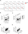
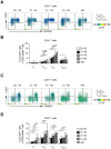
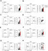
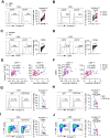
Aging is associated with immune dysregulation, especially T cell disorders, which result in increased susceptibility to various diseases. Previous studies have shown that loss of co-stimulatory receptors or accumulation of co-inhibitory molecules play important roles in T cell aging. In the present study, CD70, which was generally regarded as a costimulatory molecule, was found to be upregulated on CD4+ and CD8+ T cells of elderly individuals. Aged CD70+ T cells displayed a phenotype of over-activation, and expressed enhanced levels of numerous inhibitory receptors including PD-1, 2B4 and LAG-3. CD70+ T cells from elderly individuals exhibited increased susceptibility to apoptosis and high levels of inflammatory cytokines. Importantly, the functional dysregulation of CD70+ T cells associated with aging was reversed by blocking CD70. Collectively, this study demonstrated CD70 as a prominent regulator involved in immunosenescence, which led to defects and overwhelming inflammatory responses of T cells during aging. These findings provide a strong rationale for targeting CD70 to prevent dysregulation related to immunosenescence.
Keywords: CD70; T cell aging; co-inhibitory molecules; immunosenescence; overwhelming inflammatory responses.
Conflict of interest statement
Figures

Similar articles
-
Increased CD8+CD27+perforin+ T cells and decreased CD8+CD70+ T cells may be immune biomarkers for aplastic anemia severity.Blood Cells Mol Dis. 2019 Jul;77:34-42. doi: 10.1016/j.bcmd.2019.03.009. Epub 2019 Mar 30. Blood Cells Mol Dis. 2019. PMID: 30953940
-
T-cell Immunoglobulin and ITIM Domain Contributes to CD8+ T-cell Immunosenescence.Aging Cell. 2018 Apr;17(2):e12716. doi: 10.1111/acel.12716. Epub 2018 Jan 19. Aging Cell. 2018. PMID: 29349889 Free PMC article.
-
Increased expression of CD80, CD86 and CD70 on T cells from HIV-infected individuals upon activation in vitro: regulation by CD4+ T cells.Eur J Immunol. 1996 Aug;26(8):1700-6. doi: 10.1002/eji.1830260806. Eur J Immunol. 1996. PMID: 8765009
-
Immunosenescence: the potential role of myeloid-derived suppressor cells (MDSC) in age-related immune deficiency.Cell Mol Life Sci. 2019 May;76(10):1901-1918. doi: 10.1007/s00018-019-03048-x. Epub 2019 Feb 20. Cell Mol Life Sci. 2019. PMID: 30788516 Free PMC article. Review.
-
Benefit delayed immunosenescence by regulating CD4+T cells: A promising therapeutic target for aging-related diseases.Aging Cell. 2024 Oct;23(10):e14317. doi: 10.1111/acel.14317. Epub 2024 Aug 18. Aging Cell. 2024. PMID: 39155409 Free PMC article. Review.
Cited by
-
Phenotypic and functional alterations of monocyte subsets with aging.Immun Ageing. 2022 Dec 13;19(1):63. doi: 10.1186/s12979-022-00321-9. Immun Ageing. 2022. PMID: 36514074 Free PMC article.
-
The significance of long chain non-coding RNA signature genes in the diagnosis and management of sepsis patients, and the development of a prediction model.Front Immunol. 2024 Dec 12;15:1450014. doi: 10.3389/fimmu.2024.1450014. eCollection 2024. Front Immunol. 2024. PMID: 39735547 Free PMC article.
-
A silver bullet for ageing medicine?: clinical relevance of T-cell checkpoint receptors in normal human ageing.Front Immunol. 2024 Feb 1;15:1360141. doi: 10.3389/fimmu.2024.1360141. eCollection 2024. Front Immunol. 2024. PMID: 38361938 Free PMC article. Review.
-
Increased PD-1+ NK Cell Subset in the Older Population.Int J Gen Med. 2024 Feb 26;17:651-661. doi: 10.2147/IJGM.S452476. eCollection 2024. Int J Gen Med. 2024. PMID: 38435114 Free PMC article.
-
Associations between CD70 methylation of T cell DNA and age in adults with systemic lupus erythematosus and population controls: The Michigan Lupus Epidemiology & Surveillance (MILES) Program.J Autoimmun. 2024 Jan;142:103137. doi: 10.1016/j.jaut.2023.103137. Epub 2023 Dec 8. J Autoimmun. 2024. PMID: 38064919 Free PMC article.
References
Publication types
MeSH terms
Substances
LinkOut - more resources
Full Text Sources
Research Materials

