. 2020 Jun 19;15(6):e0232654.
doi: 10.1371/journal.pone.0232654. eCollection 2020.

Comprehensive genetic analysis of 961 unrelated Duchenne Muscular Dystrophy patients: Focus on diagnosis, prevention and therapeutic possibilities

Affiliations

Comprehensive genetic analysis of 961 unrelated Duchenne Muscular Dystrophy patients: Focus on diagnosis, prevention and therapeutic possibilities

Shalini H Kumar et al. PLoS One. .

Abstract

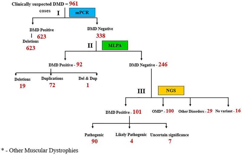
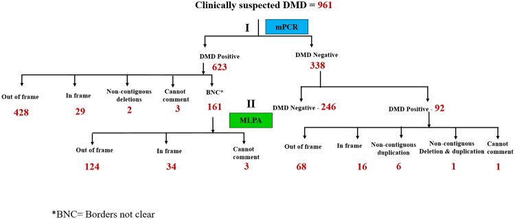
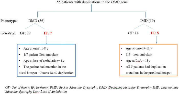
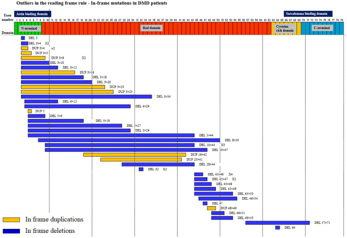
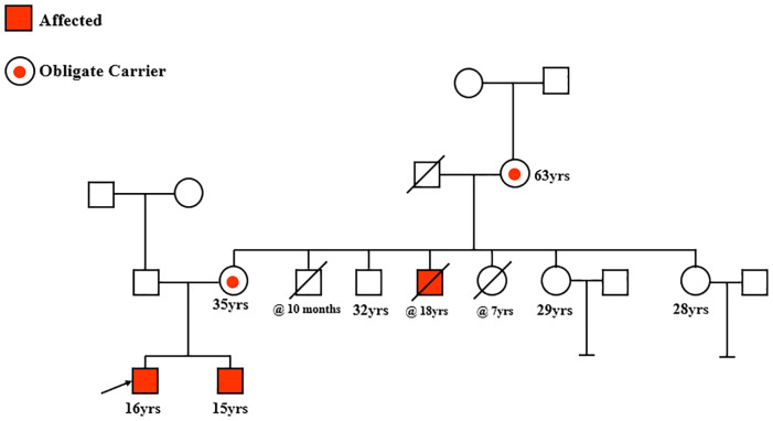
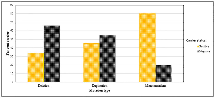
Recently DNA sequencing analysis has played a vital role in the unambiguous diagnosis of clinically suspected patients with Duchenne Muscular Dystrophy (DMD). DMD is a monogenic, X-linked, recessive, degenerative pediatric neuromuscular disorder affecting males, invariably leading to fatal cardiopulmonary failure. Early and precise diagnosis of the disease is an essential part of an effective disease management strategy as care guidelines and prevention through counseling need to be initiated at the earliest particularly since therapies are now available for a subset of patients. In this manuscript we report the DMD gene mutational profiles of 961 clinically suspected male DMD patients, 99% of whom were unrelated. We utilized a molecular diagnostic approach which is cost-effective for most patients and follows a systematic process that sequentially involves identification of hotspot deletions using mPCR, large deletions and duplications using MLPA and small insertions/ deletions and point mutations using an NGS muscular dystrophy gene panel. Pathogenic DMD gene mutations were identified in 84% of patients. Our data compared well with the frequencies and distribution of deletions and duplications reported in the DMD gene in other published studies. We also describe a number of rare in-frame mutations, which appeared to be enriched in the 5' proximal hotspot region of the DMD gene. Furthermore, we identified a family with a rare non-contiguous deletion mutation in the DMD gene where three males were affected and two females were deemed carriers. A subset of patients with mutations in the DMD gene who are likely to benefit therapeutically from new FDA and EMA approved drugs were found in our cohort. Given that the burden of care for DMD patients invariably falls on the mothers, particularly in rural India, effective genetic counseling followed by carrier screening is crucial for prevention of this disorder. We analyzed the carrier status of consented female relatives of 463 probands to gauge the percentage of patients with familial disease. Our analysis revealed 43.7% of mothers with DMD gene mutations. Our comprehensive efforts, involving complete genetic testing coupled with compassionate genetic counseling provided to DMD patients and their families, are intended to improve the quality of life of DMD patients and to empower carrier females to make informed reproductive choices to impede the propagation of this deadly disease.


Conflict of interest statement

The authors have declared that no competing interests exist.

Figures

Fig 1
Fig 1. Diagnostic strategy used to identify mutations in 961 suspected DMD patients.
DNA isolated from patient samples was subjected to the following steps: Step I involved multiplex PCR (mPCR), step II involved MLPA and step III involved NGS (Next Gen sequencing). The numbers in red indicate number of patients. OMD: Other muscular dystrophies.
Fig 2
Fig 2. Stratification of DMD positive patients based on mutation type.
Fig 3
Fig 3. Deletion mutations in the DMD gene in 642 patients mapped to two hotspot regions: Proximal (exons 1–22) and distal (exons 43–55).
The frequency of deletion mutations in the DMD gene has been shown. Blue bars represent patients with deletions beginning in the indicated exon. Red bars represent patients with deletion mutations ending in the indicated exon.
Fig 4
Fig 4. An overview of the genotype: Phenotype correlate of patients with deletions mutations in the DMD gene.
Patient numbers are indicated. The number of patients defying the reading frame rule are marked in red. OF: out of frame; IF In-frame; BMD: Becker Muscular Dystrophy; IMD: Intermediate Muscular Dystrophy; DMD: Duchenne Muscular Dystrophy; LoA: Loss of ambulation.
Fig 5
Fig 5. Genotype: Phenotype overview of patients with duplication mutations in the DMD gene.
The number of patients in each segment has been numerically indicated. The number of patients defying the reading frame rule is marked in red. OF: out of frame; IF In-frame; BMD: Becker’s Muscular Dystrophy; IMD: Intermediate Muscular Dystrophy; DMD: Duchenne Muscular Dystrophy; LoA: Loss of ambulation.
Fig 6
Fig 6. A summary of in-frame mutations in DMD patients shows an enrichment of mutations arising in the proximal DMD gene hotspot.
50 in-frame deletion and duplication mutations in DMD patients were mapped to the 79 exons of the DMD gene, which is depicted with its functional domains: N-terminus, Rod domains, Cysteine rich domain and C-terminus. The precise mutation in each case and its frequency (where applicable) has been indicated. In-frame duplications are in yellow. In-frame deletions are in blue.
Fig 7
Fig 7. Pedigree of a family with a rare non-contiguous deletion mutation in the DMD gene: Del exons 45–50 and 53–54.
Fig 8
Fig 8. Carrier frequencies based on type of mutations in the DMD gene.
Deletions, duplications and micro (point) mutations occurring in both carrier (positive) and normal (negative) mothers was assessed. Point mutations were most frequent in carrier mothers.

References

    1. Muntoni F, Torelli S, Ferlini A. Dystrophin and mutations: One gene, several proteins, multiple phenotypes. Lancet Neurology. 2003. 10.1016/S1474-4422(03)00585-4 - DOI - PubMed
    1. Moat SJ, Bradley DM, Salmon R, Clarke A, Hartley L. Newborn bloodspot screening for Duchenne Muscular Dystrophy: 21 years experience in Wales (UK). Eur J Hum Genet. 2013. 10.1038/ejhg.2012.301 - DOI - PMC - PubMed
    1. Deconinck N, Dan B. Pathophysiology of Duchenne Muscular Dystrophy: Current Hypotheses. Pediatric Neurology. 2007. 10.1016/j.pediatrneurol.2006.09.016 - DOI - PubMed
    1. McDonald CM, Henricson EK, Abresch RT, Duong T, Joyce NC, Hu F, et al. Long-term effects of glucocorticoids on function, quality of life, and survival in patients with Duchenne muscular dystrophy: a prospective cohort study. Lancet. 2018. 10.1016/S0140-6736(17)32160-8 - DOI - PubMed
    1. Hoffman EP, Brown RH, Kunkel LM. Dystrophin: The protein product of the duchenne muscular dystrophy locus. Cell. 1987. 10.1016/0092-8674(87)90579-4 - DOI - PubMed