. 2020 Jun 19;15(6):e0234873.
doi: 10.1371/journal.pone.0234873. eCollection 2020.

Two ways of epigenetic silencing of TFPI2 in cervical cancer

Affiliations

Two ways of epigenetic silencing of TFPI2 in cervical cancer

Alexandra Fullár et al. PLoS One. .

Abstract

Objective: Comparison of human mRNA microarray results from tumor-associated and normal cervical fibroblasts revealed significant TFPI2 downregulation in tumor-associated fibroblasts isolated from cervical cancer, indicating that TFPI2 downregulation may play an important role in the pathogenesis of the disease. In the present work, we investigated the mechanism of TFPI2 downregulation in tumor-associated fibroblasts and tumor cells.

Methods: In vitro models of monocultures and co-cultures were established with tumor cells and fibroblasts to explore the changes of TFPI-2 expression and epigenetic modifications of the TFPI2 gene.

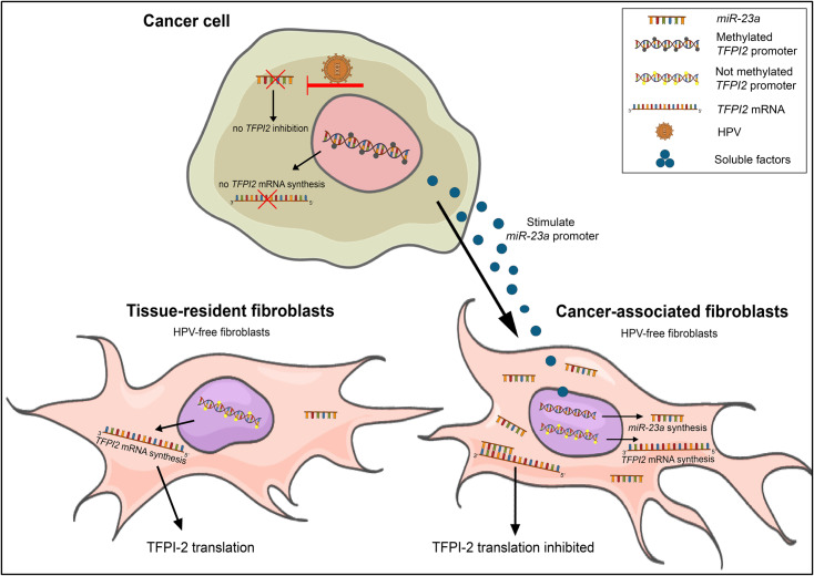
Results: The TFPI2 gene was hypermethylated only in tumor cells. Reduction of TFPI-2 protein levels in tumor-associated fibroblasts, although the gene was not methylated, suggested alternative regulatory mechanisms of gene expression, such as inhibition by microRNAs. The expression pattern of miR-23a, a gene thought to inhibit TFPI2 translation, showed changes strongly correlated to detected TFPI-2 protein alterations. Transfections with miR-23a mimics resulted in a decrease of TFPI-2 protein expression whereas miR-23a inhibitors increased the TFPI-2 amount. Due to downregulation of miR-23a expression by HPV in cancer cells, TFPI2 was silenced by promoter methylation. In contrary, miR-23a was active in HPV-free fibroblasts and inactivated TFPI2.

Conclusion: These results indicate dual epigenetic inhibition of TFPI2 on the transcription level by promoter methylation in cancer cells and on the translation level by miR-23a in tumor-associated fibroblasts. As a consequence, inactivation of the TFPI2 gene plays a strategic role in the progression of cervical cancer.


Conflict of interest statement

The employment of author (ZP) at by Maternity Obstetrics and Gynecology Private Clinic does not alter our adherence to PLOS ONE policies on sharing data and materials. The remaining authors have no competing interests to disclose.

Figures

Fig 1
Fig 1. Expression of TFPI2 in fibroblasts and cancer cells.
Among 67 significantly changed mRNAs, TFPI2 and its precursor in TF were most significantly downregulated compared to that in NF cells (panel A). The human mRNA microarray was validated by real-time PCR on cell cultures derived from explants of case 2 (S1 Table) and on C cell line. The mRNA levels of TFPI2 changed 0.01-fold (Student’s t-test: p < 0.001) in TF and 0.23-fold (Student’s t-test: p < 0.001) in MF compared to NF. TFPI2 was not expressed either in T or in C (panel B). Stars indicate significance: **p < 0.01. Results were expressed as mean, error bars represent SD (n = 3).
Fig 2
Fig 2. Morphology of the cells used for the experiments and typical co-cultures.
In one of the tumor-associated fibroblast cultures (TF) a few vimentin and cytokeratin double positive cells were observed. These TF-like cells (marked with white star) corresponded to EMT transformed cancer cells. These newly formed cells started to proliferate and displaced the real fibroblasts from the culture. They lost their vimentin positivity and transformed into round shape cytokeratin positive T cells (marked with white arrow on MET picture). MET: mesenchymal-epithelial transition, EMT: epithelial-mesenchymal transition. The representative images show 200x magnification with scale bar of 100 μm, and 100x magnification with scale bar of 50 μm (panel A). In direct co-cultures both types of tumor cells (T and C) exhibited cytokeratin positivity, while TFs were positive for vimentin (TF+T: scale bar is 100 μm; and TF+C: scale bar is 60 μm). T represents real tumor cells similar to CSCC7 (C) cells (panel B). In the H&E stained co-cultures T and the C cells were grown with NF, TF and MF (black stars), respectively. The morphology and the arrangement of the two types of cancer cells (T and C, marked with black arrows) are comparable. They have similar epithelial morphology forming nests. All types of fibroblasts display spindle-like morphology, with oval nuclei but they have small differences: NF (normal fibroblasts) cells are thin and long; TF cells are wider with more cytoplasm, while MF (fibroblasts form lymph node metastasis) cells are the biggest cells among the fibroblasts. The representative images show 200x magnifications, scale bar of 100 μm (panels C and D). Red: cytokeratin, green: vimentin.
Fig 3
Fig 3. Expression of TFPI-2 protein.
Western blot of TFPI-2 protein showing 33, 31, and 27 kDa bands in fibroblasts, cancer cells and their co-cultures, respectively (panels A-F). Decreasing levels of TFPI-2 protein expression was detected in fibroblasts (F) from NF, through MF to TF. Similarly to the two tumor cell cultures (T, C) the expression was hardly detectable in TF (panels A and B). Direct contact of NF or MF with tumor cells downregulated the expression of TFPI-2 (panels C-F). Thus, the final densitometric value in co-cultures was lower than the sum of densitometry in 2 monocultures (sum of monocultures F + T or F + C). ∑: sum. Results were expressed as mean, error bars represent SD (n = 2).
Fig 4
Fig 4. TFPI-2 immunohistochemistry of cervical cancer specimens.
TFPI-2 was detected on the epithelial layer of normal cervical area. In addition to the presence in the nucleus, modest cytoplasmic positivity was observed (A). TFPI-2 was detected proximal to the cervical surface cytoplasm of tumorous nest and in lower amounts in the cytoplasm of fibroblasts (B-E). However, the deeper area of the specimens, both tumor and the surrounding connective tissue, lacked TFPI-2 staining (F). The nuclei were counterstained with hematoxylin blue. E: epithelial layer, S: stroma, T: tumor, black arrows: plasma cells. The representative images show 200x magnifications with, scale bar set to 100 μm.
Fig 5
Fig 5. TFPI2 gene methylation.
In assays 4 and 5 for TFPI2 MS-HRM melting curves of cervical fibroblasts and cervical tumor cells (red line) were compared to the methylation standards (green lines with different degree of methylation: 100%, 75%, 50%, 25%, 0%) displaying characteristic melting profiles. These showed evidence of hypermethylation only in tumor cells and hypomethylation in fibroblasts both in monocultures and in indirect co-cultures (panels A-C). Differentially methylated CpG sites of TFPI2 region in assay 6 for cervical fibroblast and tumor cells using methylation pyrosequencing were visualized on the heat map. Both tumor cultures (T, C) showed higher methylation levels in all CpG sites, whereas the methylation level was variable in fibroblasts. Fibroblasts were not, or hardly, methylated at CpG_4 site, indicating the importance of this region in promoter silencing (panel D). Green = 100% methylated; Red = 0% methylated.
Fig 6
Fig 6. Expression of miRNAs targeting TFPI2 in cervical cell cultures/lines.
Expression of miRNAs predicted to inhibit TFPI2 translation. Compared to NF, TF cells (black column) showed increased expression of miR-616-3p, miR-646 and miR-23a. The changes of miR-23a corresponded the highest level of TFPI-2 alteration in various cell cultures (n = 3) (panel A). For validation the role of miR-23a, its mimics and inhibitors were transfected into NF cells. Comparing to their negative control (NC) the mimics decreased and the inhibitors increased the expression of TFPI-2 supporting the significance of miR-23a in the regulation of the protein. Black line: pictures were edited to remove one irrelevant lane (panel B). Densitometry indicated that miR-23a-3p affects mostly TFPI-2 protein expression. UT: untreated NF used as positive control (n = 2) (panel C). Results were expressed as mean, error bars represent SD.
Fig 7
Fig 7. HPV status determination by HPV E6 ORF-specific nested PCR on DNAs isolated from cervical cancers.
Fibroblasts (NF, TF) are HPV-free cells, and the HPV16 positivity of C tumor cells was confirmed (panel A). FFPE sample of the second case and its tumor cell culture (T) showed the presence of HPV16 (panel B). The size of PCR fragments corresponds to that of the size as the positive control. NC: negative control of first (no DNA) and second round of nested PCR; HPV16 DNA: cloned plasmid containing HPV16 DNA (positive control). The values 1x, 10x and 100x represent different dilutions.
Fig 8
Fig 8. Two ways of epigenetic silencing of TFPI2 in cervical cancer.
Continuous E6/E7 expression of HPV hinders miR-23a in cancer cells, hereby the TFPI-2 could be expressed. However, this is overridden by promoter methylation epigenetic silencing thus finally TFPI-2 expression is inhibited in cancer cells. In spite of that TFPI2 promoter was not methylated in fibroblasts, TFs did not produce the protein. This is explained by the elevated intracellular concentrations of miR-23a stimulated by a so far unknown factor(s) of cancer cells. Translation of TFPI2 is inhibited by binding of miR-23a to the 3’ UTR of TFPI2 mRNA.

References

    1. Walboomers JM, Jacobs MV, Manos MM, Bosch FX, Kummer JA, Shah KV, et al. Human papillomavirus is a necessary cause of invasive cervical cancer worldwide. J Pathol. 1999;189(1):12–9. Epub 1999/08/19. 10.1002/(SICI)1096-9896(199909)189:1<12::AID-PATH431>3.0.CO;2-F . - DOI - PubMed
    1. Fullar A, Dudas J, Olah L, Hollosi P, Papp Z, Sobel G, et al. Remodeling of extracellular matrix by normal and tumor-associated fibroblasts promotes cervical cancer progression. BMC Cancer. 2015;15:256 Epub 2015/04/18. 10.1186/s12885-015-1272-3 - DOI - PMC - PubMed
    1. Lorusso G, Rüegg C. The tumor microenvironment and its contribution to tumor evolution toward metastasis. Histochemistry and Cell Biology. 2008;130(6):1091–103. 10.1007/s00418-008-0530-8 - DOI - PubMed
    1. Kalluri R, Zeisberg M. Fibroblasts in cancer. Nat Rev Cancer. 2006;6(5):392–401. Epub 2006/03/31. 10.1038/nrc1877 . - DOI - PubMed
    1. Fullar A, Kovalszky I, Bitsche M, Romani A, Schartinger VH, Sprinzl GM, et al. Tumor cell and carcinoma-associated fibroblast interaction regulates matrix metalloproteinases and their inhibitors in oral squamous cell carcinoma. Exp Cell Res. 2012;318(13):1517–27. Epub 2012/04/21. 10.1016/j.yexcr.2012.03.023 - DOI - PMC - PubMed

Publication types