. 2020 Aug 4;32(2):229-242.e8.
doi: 10.1016/j.cmet.2020.06.002. Epub 2020 Jun 18.

Targeting DGAT1 Ameliorates Glioblastoma by Increasing Fat Catabolism and Oxidative Stress

Affiliations

Targeting DGAT1 Ameliorates Glioblastoma by Increasing Fat Catabolism and Oxidative Stress

Xiang Cheng et al. Cell Metab. .

Abstract

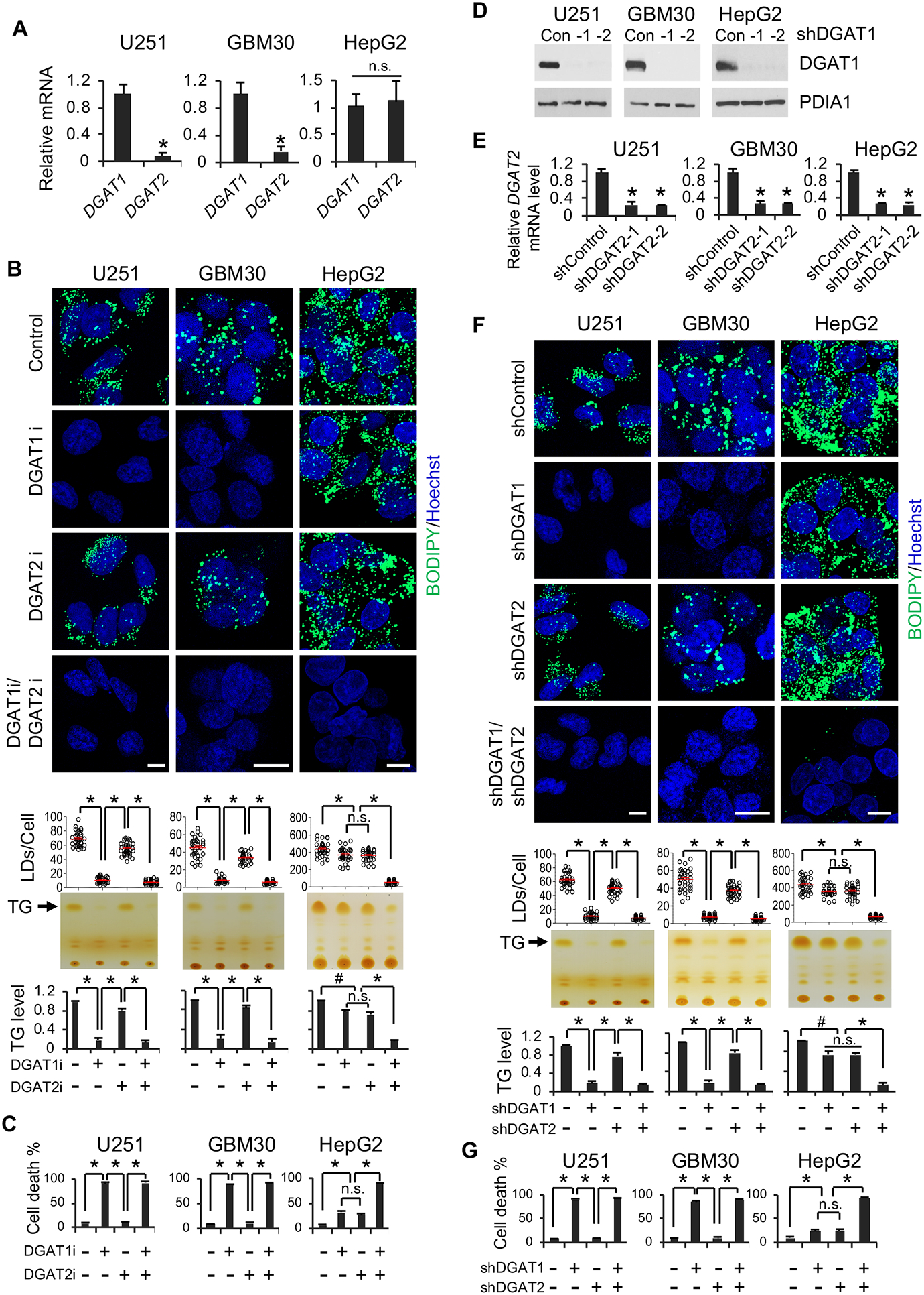
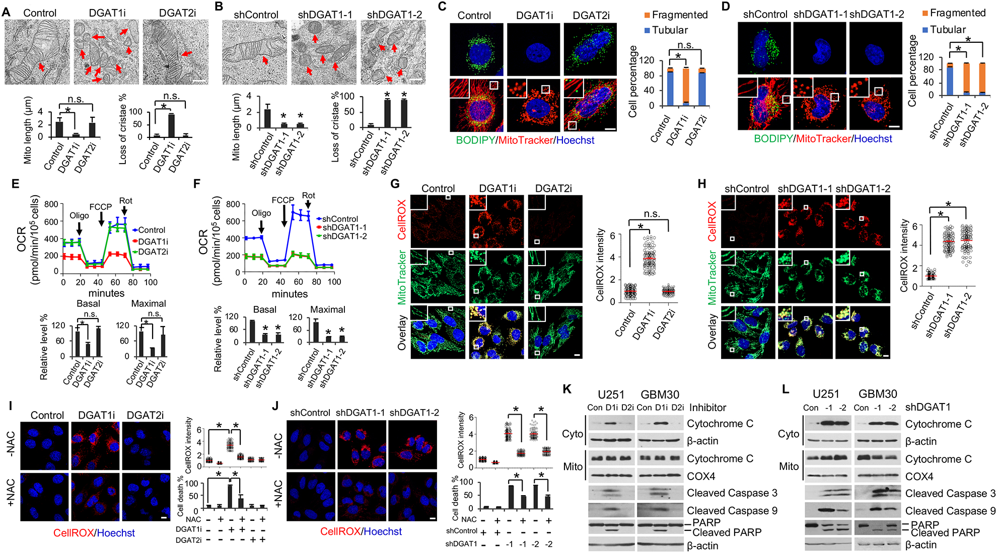
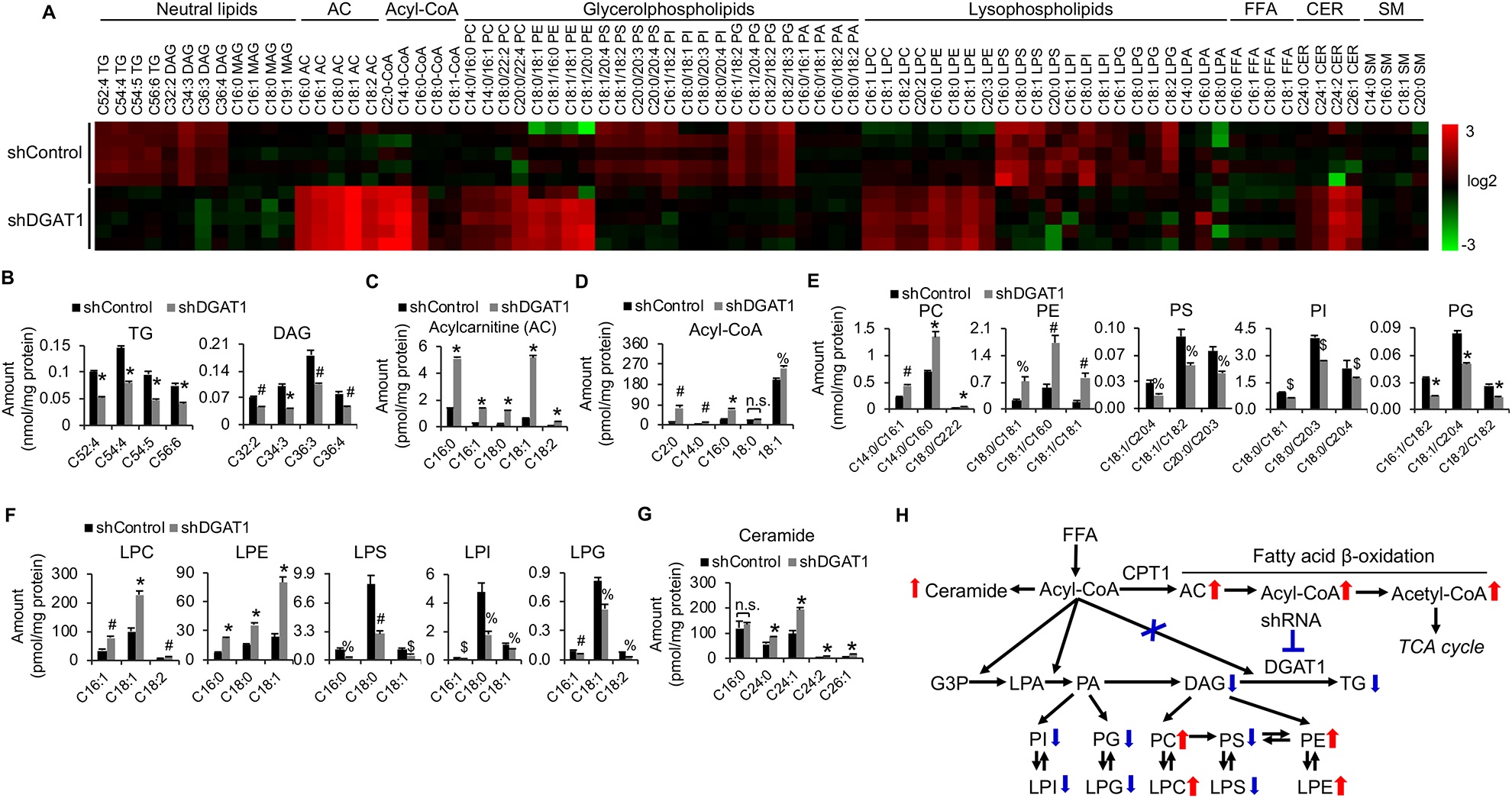
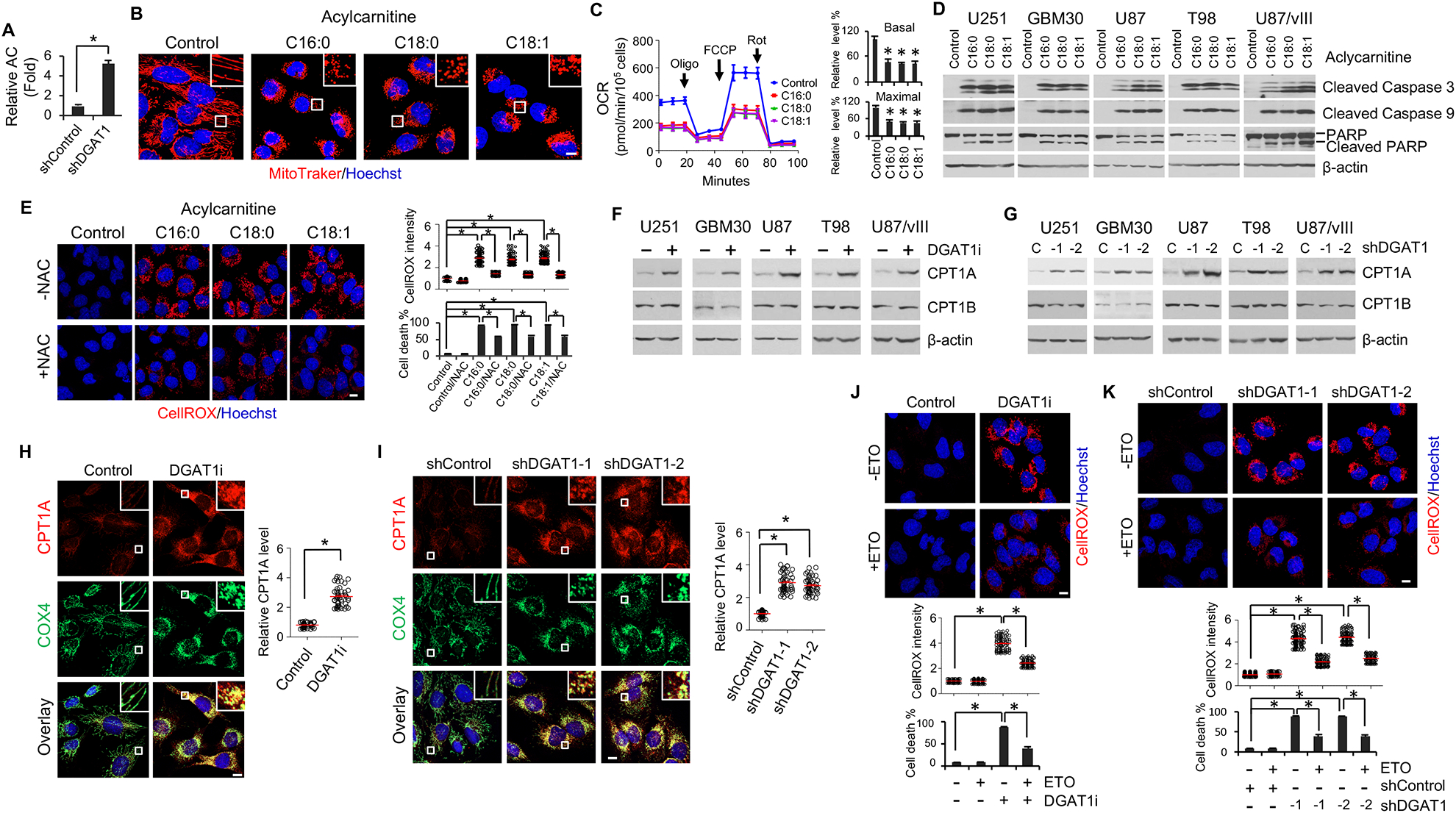
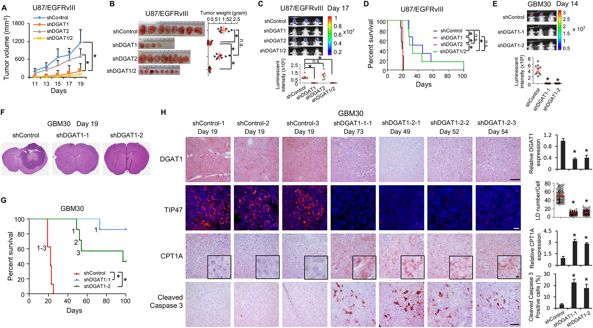
Glioblastoma (GBM), a mostly lethal brain tumor, acquires large amounts of free fatty acids (FAs) to promote cell growth. But how the cancer avoids lipotoxicity is unknown. Here, we identify that GBM upregulates diacylglycerol-acyltransferase 1 (DGAT1) to store excess FAs into triglycerides and lipid droplets. Inhibiting DGAT1 disrupted lipid homeostasis and resulted in excessive FAs moving into mitochondria for oxidation, leading to the generation of high levels of reactive oxygen species (ROS), mitochondrial damage, cytochrome c release, and apoptosis. Adding N-acetyl-cysteine or inhibiting FA shuttling into mitochondria decreased ROS and cell death induced by DGAT1 inhibition. We show in xenograft models that targeting DGAT1 blocked lipid droplet formation, induced tumor cell apoptosis, and markedly suppressed GBM growth. Together, our study demonstrates that DGAT1 upregulation protects GBM from oxidative damage and maintains lipid homeostasis by facilitating storage of excess FAs. Targeting DGAT1 could be a promising therapeutic approach for GBM.

Keywords: DGAT1; ROS; acylcarnitine; fatty acids; glioblastoma; lipid droplets; lipotoxicity; mitochondria; oxidative stress; triglycerides.


Conflict of interest statement

Declaration of Interests The authors declare that there are no competing interests.

Figures

Figure 1.
Figure 1.. GBM tumors contain large amounts of TGs and express high levels of DGAT1 that are associated with poor patient survival.
(A) TLC analysis of TG levels in human normal brains vs. tumor tissues from individuals with GBM. *P < 0.001. (B) A representative Western blot (n = 2 blots in total) of DGAT1 from human normal brain vs. GBM tumors. Protein disulfide-isomerase family A, member 1 (PDIA1), an ER-resident protein, was used as a loading control. (C) Representative IHC staining (n = 3 images in total) of DGAT1 in human normal brain vs. GBM tumor samples (upper panels). IF staining (n = 5 images in total) of LDs via using TIP47 antibody (lower panels). Nucleus was stained with DAPI. Scale bar, 50 μm for IHC, 10 μm for IF images. (D) RT-qPCR analysis of mRNA expression in human GBM tumor samples (n = 10) and normalized to DGAT1 average expression. *P < 0.001. (E) Boxplot analysis of gene expression in samples from individuals with GBM (n = 153), ovarian (n = 303), prostate (n = 497), breast (n = 1009) and liver (n = 371) cancer in the TCGA RNA-seq databases. RPKM, reads per kilobase million. *P < 0.001. (F and G) IHC analysis of DGAT1 expression in glioma tissues in the TMA (n = 62) (F, upper panels). LDs were detected by IF via TIP47 staining (red) (F, lower panels). Scale bar, 20 μm for IHC, 10 μm for IF. DGAT1 levels were quantified by H-score (G). *P < 0.01. PA, pilocytic astrocytoma, grade I; A2, astrocytoma grade II; AA, anaplastic astrocytoma, grade III. (H and I) Kaplan-Meier plot of survival data from individuals with GBM based on DGAT1 protein levels in TMA analyzed in panels F and G (mean = 180) (H), or based on DGAT1 mRNA levels in GBM TCGA database (RNA-seq) (I). The optimal cut-off 9.503 was applied to stratify the high vs. low groups. See also Figure S1.
Figure 2.
Figure 2.. Inhibition of DGAT1, but not DGAT2, significantly suppresses TG and LD formation and induces GBM cell death.
(A) RT-qPCR analysis of mRNA expression (mean ± SD, n = 3) in GBM cells (U251 and GBM30) and in liver cancer cell line (HepG2). *P < 0.01; n.s., not significant. (B) Representative fluorescence imaging (n = 6 images in total) of LDs stained with BODIPY 493/503 (green) (upper panels) and TLC analysis (n = 3) of TG levels (lower panels) in different cancer cells treated with/without DGAT1 inhibitor A-922500 (20 μg/ml) or DGAT2 inhibitor PF-06424439 (20 μg/ml) for 24 hr. Nuclei were stained with Hoechst 33342 (blue). Scale bar, 10 μm. *P < 0.001; #P < 0.05. (C) Percentage of dead cells (mean ± SD, n = 3) after treatment with DGAT1 or DGAT2 inhibitor for 3 days as in panel B. *P < 0.0001. (D) A representative Western blot (n = 3 blots in total) of DGAT1 expression after shRNA knockdown for 48 hr. (E) RT-qPCR analysis of DGAT2 mRNA expression (mean ± SD, n = 3) in different cancer cells after shRNA knockdown for 48 hr. *P < 0.01. (F) Representative fluorescence imaging (n = 6 images in total) of LDs stained as panel B (upper panels) and TLC analysis of TG levels (lower panels) in different cancer cells after shRNA knockdown of DGAT1 or DGAT2 for 48 hr. Scale bar, 10 μm. *P < 0.001; #P < 0.05. (G) Percentage of dead cells (mean ± SD, n = 3) after shRNA knockdown of DGAT1 or DGAT2 for 4 days. *P < 0.0001; #P < 0.001. See also Figure S2.
Figure 3.
Figure 3.. Inhibition of DGAT1 causes mitochondrial damage, ROS production and GBM cell apoptosis.
(A and B) Representative TEM imaging (n = 20 images in total) of the mitochondria in U251 cells treated with/without DGAT1 inhibitor A-922500 (20 μg/ml) or DGAT2 inhibitor PF-06424439 (20 μg/ml) for 24 hr (A) or shDGAT1 knockdown for 48 hr (B). Scale bar, 500 nm. Red arrows indicate mitochondria. Over 100 mitochondria in each group were quantified (mean ± SEM). *P < 0.0001. (C and D) Representative fluorescence imaging (n = 12 images in total) of LDs (green) and mitochondria (red) in U251 cells treated with/without DGAT1 or DGAT2 inhibitor (C) or shDGAT1 (D) as in panels A and B. Scale bar, 10 μm. More than 100 cells were quantified for tubular or fragmented mitochondria (mean ± SEM). *P < 0.001. (E and F) Oxygen consumption rate (OCR) (mean ± SD, n = 3) in U251 cells treated with/without DGAT1 or DGAT2 inhibitor (E) or shRNA (F) as in panels A and B. Oligo, oligomycin; FCCP, carbonyl cyanide 4-trifluoromethoxy-phenylhydrazone; Rot, rotenone. *P < 0.0001. (G and H) Representative fluorescence imaging (n = 12 images in total) of ROS (red) detected with CellROX Deep Red and mitochondria (green) in U251 cells treated with/without DGAT1 or DGAT2 inhibitor (G) or shRNA (H) as in panels A and B. ROS levels were quantified in more than 100 cells (mean ± SD). *P < 0.0001. Scale bar, 10 μm. (I and J) Representative fluorescence images (n = 12 images in total) of ROS in U251 cells treated with/without DGAT1 inhibitor (I) or shRNA (J) as in panels A and B in the presence or absence of NAC (1 mM). Scale bar, 10 μm. Dead cell percentage were counted after treatment for 3 days (mean ± SD, n = 3) (right bottom panels). *P < 0.0001. (K and L) A representative Western blot (n = 3 blots in total) of GBM cells treated with/without DGAT1 (D1i) inhibitor A-922500 (20 μg/ml) or DGAT2 (D2i) inhibitor PF-06424439 (20 μg/ml) for 24 hr (K) or shRNA against DGAT1 for 72 hr (L). Cyto, cytosol. Mito, mitochondria. See also Figures S3, S4 and S5.
Figure 4.
Figure 4.. Genetic inhibition of DGAT1 significantly alters lipid homeostasis and dramatically elevates acylcarnitine and acetyl-CoA levels in GBM cells.
(A) Heatmap of representative lipids in U251 cells with/without shRNA knockdown of DGAT1 (60 hr) analyzed by lipidomics. AC, acylcarnitine; FFA, free fatty acid; CER, ceramide; SM, sphingomyelin. (B-G) Levels of representative individual lipid species in U251 cells with/without shDGAT1 knockdown (mean ± SEM, n = 5). *P < 0.0001; #P < 0.001; %P < 0.01; $P < 0.05 in comparison to shControl cells. PC, phosphatidylcholine; PE, phosphatidylethanolamine; PS, phosphatidylserine; PI, phosphatidylinositol; PG phosphatidylglycerol; PA, phosphatidic acid; LPC, lysophosphatidylcholine; LPE, lysophosphatidylethanolamine; LPS, lysophosphatidylserine; LPI, lysophosphatidylinositol; LPG, lysophosphatidylglycerol; LPA, lysophosphatidic acid. (H) Summary of the lipid profiling changes in U251 cells after knockdown of DGAT1. G3P, glycerol-3-phosphate. See also Figure S6.
Figure 5.
Figure 5.. Elevated acylcarnitine induced by DGAT1 inhibition damages the mitochondria and are associated with CPT1A upregulation.
(A) Relative total acylcarnitine (AC) level in U251 cells with/without DGAT1 knockdown (mean ± SEM, n = 5). *P < 0.0001. (B-D) Representative fluorescence imaging (n = 6 images in total) of mitochondria stained with MitoTracker Red (red) (B), OCR measurement (mean ± SD, n = 3) (C) or Western blot analysis (n = 3 blots in total) of apoptosis markers (D) in U251 cells treated with different acylcarnitines (20 μM) or vehicle (ethanol) as control for 24 hr. Nuclei were stained with Hoechst 33342 (blue). Scale bar, 10 μm. *P < 0.0001 as compared with control. (E) Representative fluorescence imaging (n = 12 images in total) of ROS (left) by CellROX staining (red) and its quantification (right) (mean ± SEM, n = 100 cells) in U251 cells treated with acylcarnitines (20 μM) for 24 hr in the presence or absence of NAC (1 mM). Scale bar, 10 μm. Cell death was determined after 2 days of treatment (mean ± SD, n = 3). *P < 0.001. (F-I) A representative Western blot (n = 3 blots in total) (F and G) or IF (n = 12 images in total) (H and I) analysis of CPT1A expression levels in U251 cells upon pharmacological inhibition (A-922500, 20 μg/ml for 24 hr) or shRNA knockdown (48 hr) of DGAT1. Cytochrome c oxidase subunit 4 (COX4) was stained by IF (green) to show mitochondria (H and I). CPT1A level was quantified in more than 50 cells (mean ± SEM) (H and I). *P < 0.0001. (J and K) Representative fluorescence imaging (n = 12 images in total) and quantification of ROS (mean ± SEM, n = 100 cells) in U251 cells treated with DGAT1 inhibitor A-922500 (20 μg/ml) for 24 hr (J) or shRNA for 48 hr (K) in the presence or absence of the CPT1 inhibitor etomoxir (ETO, 6 μM). Scale bar, 10 μm. *P < 0.0001. Cell death was quantified after 72 hr of inhibitor treatment (J) or 96 hr of shRNA knockdown (K) (mean ± SD, n = 3). See also Figure S7.
Figure 6.
Figure 6.. Genetic inhibition of DGAT1 significantly suppresses tumor growth and prolongs overall survival in GBM-bearing mice
(A-D) The effects of knockdown of DGAT1 or DGAT2 in U87/EGFRvIII cells-derived subcutaneous (A and B) or intracranial (C and D) tumor models. Tumor growth in mouse brain was analyzed by bioluminescence imaging at day 17 post implantation (mean ± SEM, n = 7) (C). Mouse survival was assessed by Kaplan–Meier curves (D). *P < 0.0001. (E-G) The effects of DGAT1 knockdown in primary GBM30-luciferase cells-derived intracranial mouse model. Tumor growth was analyzed by bioluminescence imaging at day 14 post implantation (mean ± SEM, n = 10) (E). Sections of mouse brain (n = 3) from day 19 post implantation were stained with H&E (F). Kaplan-Meier plot for analysis of mouse survival (n = 7)(G). The ‘1’, ‘2’ and ‘3’ in the panel G means the number of mice that reached to mortality stage and sacrificed for analysis in panel H. *P < 0.001. (H) Representative imaging (n = 5 in total images) of IHC of DGAT1, CPT1A and cleaved Caspase 3 or IF of LDs via staining TIP47 in tumor tissues from the mice indicated in panel G and sacrificed at indicated days. Five separate areas from each tumor were quantified (mean ± SEM). Scale bars, 40 μm for IHC, 10 μm for IF. *P < 0.01 See also Figure S8.
Figure 7.
Figure 7.. Pharmacological inhibition of DGAT1 dramatically suppresses GBM tumor growth and induces tumor cell apoptosis
(A-C) The effects of DGAT1 inhibitor A-900225 (120 mg/kg/day, oral gavage) in U87/EGFRvIII- or GBM30-derived subcutaneous models (n = 6) (A and B). Tumor tissues were analyzed for LDs by IF (TIP47 staining), CPT1A, and cleaved Caspase 3 levels by IHC (representative imaging from 5 images in each staining) (C). Scale bars, 10 μm for IF, 40 μm for IHC. Quantification was performed by analyzing 5 separate areas in each tumor (mean ± SEM). *P < 0.01. (D) Schematic model illustrating the function of DGAT1 in regulating lipid homeostasis and the cytotoxic effects resulting from its inhibition in GBM cells. See also Figure S9.

References

    1. Accioly MT, Pacheco P, Maya-Monteiro CM, Carrossini N, Robbs BK, Oliveira SS, Kaufmann C, Morgado-Diaz JA, Bozza PT, and Viola JP (2008). Lipid bodies are reservoirs of cyclooxygenase-2 and sites of prostaglandin-E2 synthesis in colon cancer cells. Cancer Res 68, 1732–1740. - PubMed
    1. Ackerman D, Tumanov S, Qiu B, Michalopoulou E, Spata M, Azzam A, Xie H, Simon MC, and Kamphorst JJ (2018). Triglycerides Promote Lipid Homeostasis during Hypoxic Stress by Balancing Fatty Acid Saturation. Cell Rep 24, 2596–2605 e2595. - PMC - PubMed
    1. Aruoma OI, Halliwell B, Hoey BM, and Butler J (1989). The antioxidant action of N-acetylcysteine: its reaction with hydrogen peroxide, hydroxyl radical, superoxide, and hypochlorous acid. Free Radic Biol Med 6, 593–597. - PubMed
    1. Atkins C, Liu Q, Minthorn E, Zhang SY, Figueroa DJ, Moss K, Stanley TB, Sanders B, Goetz A, Gaul N, et al. (2013). Characterization of a novel PERK kinase inhibitor with antitumor and antiangiogenic activity. Cancer Res 73, 1993–2002. - PubMed
    1. Carracedo A, Cantley LC, and Pandolfi PP (2013). Cancer metabolism: fatty acid oxidation in the limelight. Nat Rev Cancer 13, 227–232. - PMC - PubMed

Publication types