. 2020 Aug 17;30(16):3223-3230.e4.
doi: 10.1016/j.cub.2020.05.072. Epub 2020 Jun 18.

Comparative Development of the Ant Chemosensory System

Affiliations

Comparative Development of the Ant Chemosensory System

Anna R Ryba et al. Curr Biol. .

Abstract

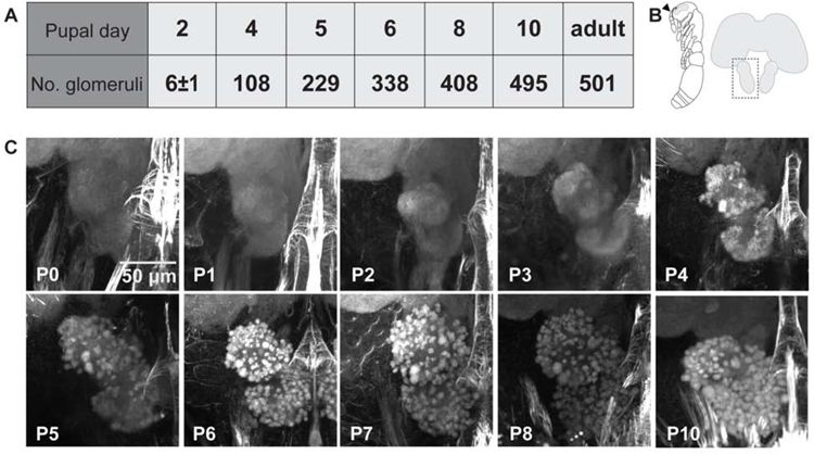
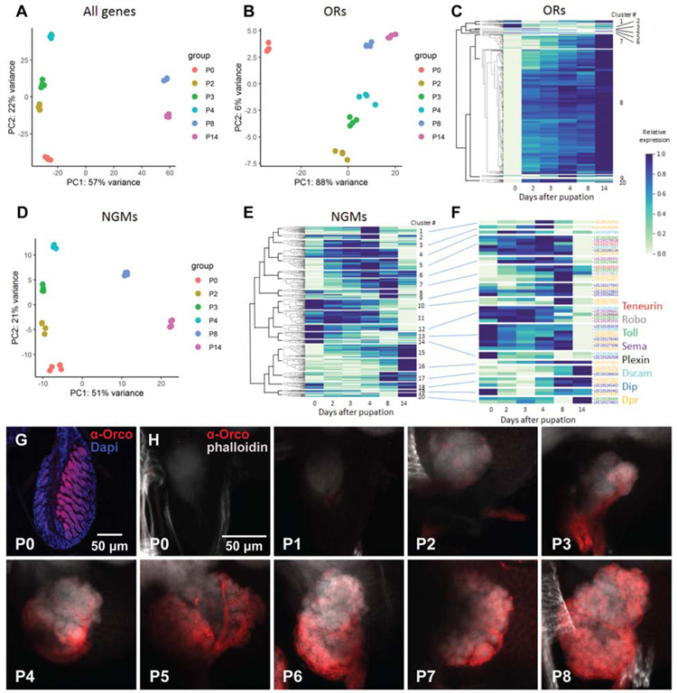
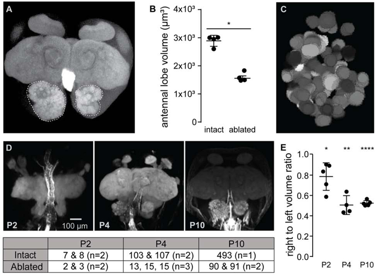
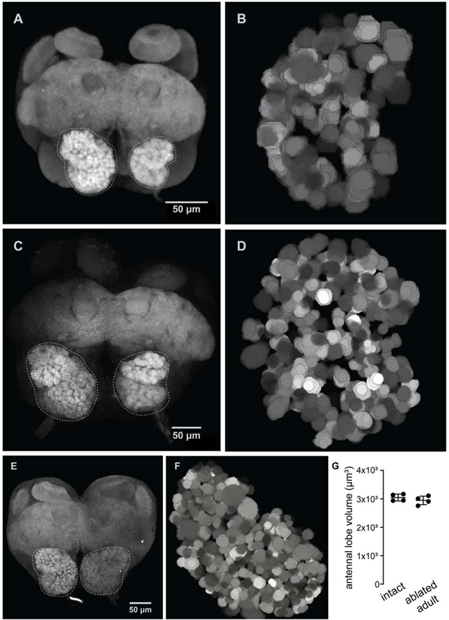
The insect antennal lobe (AL) contains the first synapses of the olfactory system, where olfactory sensory neurons (OSNs) contact second-order projection neurons (PNs). In Drosophila melanogaster, OSNs expressing specific receptor genes send stereotyped projections to one or two of about 50 morphologically defined glomeruli [1-3]. The mechanisms for this precise matching between OSNs and PNs have been studied extensively in D. melanogaster, where development is deterministic and independent of neural activity [4-6]. However, a number of insect lineages, most notably the ants, have receptor gene repertoires many times larger than D. melanogaster and exhibit more structurally complex antennal lobes [7-12]. Moreover, perturbation of OSN function via knockout of the odorant receptor (OR) co-receptor, Orco, results in drastic AL reductions in ants [13, 14], but not in Drosophila [15]. Here, we characterize AL development in the clonal raider ant, Ooceraea biroi. We find that, unlike in Drosophila, ORs and Orco are expressed before the onset of glomerulus formation, and Orco protein is trafficked to developing axon terminals, raising the possibility that ORs play a role during ant AL development. Additionally, ablating ant antennae at the onset of pupation results in AL defects that recapitulate the Orco mutant phenotype. Thus, early loss of functional OSN innervation reveals latent structure in the AL that develops independently of peripheral input, suggesting that the AL is initially pre-patterned and later refined in an OSN-dependent manner. This two-step process might increase developmental flexibility and thereby facilitate the rapid evolution and expansion of the ant chemosensory system.

Keywords: Formicidae; Ooceraea biroi; antennal lobe; chemosensation; clonal raider ant; odorant receptors; olfaction; pheromones; sensory systems; social insects.


Conflict of interest statement

Declaration of Interests The authors declare no competing interests.

Figures

Figure 1:
Figure 1:. Antennal lobe development over the first 10 days of pupation.
(A) Manual counts of glomeruli from confocal stacks of pupal brains stained with phalloidin. Glomeruli were counted in the right antennal lobe from each brain. P2 n=6 brains with average 6±1 SD glomeruli; n=1 for all other pupal time-points; adult n=2 brains with 493 and 509 glomeruli (average 501; adult counts were previously published[13]). (B) Illustration of an ant pupa with the antenna marked by an arrowhead (left), and schematic of an ant brain with the left antennal lobe boxed to show the orientation of images in (C). (C) Representative projections of confocal z-stacks through the right antennal lobe of phalloidin stained pupal brains (see also Figure S1A).
Figure 2:
Figure 2:. RNA-seq and Orco staining during pupal development.
(A) PCA of antennal gene expression for all genes at each developmental time-point. (B) PCA of odorant receptor (OR) gene expression in the antennae over the course of development (see also Figure S1B–D and Table S1). (C) Gene expression heatmap and clustering of OR genes throughout development. (D) PCA of neuronal guidance molecule (NGM) gene expression in antennae over the course of development. (E) Gene expression heatmap and clustering of NGM genes throughout development. (F) Gene expression heatmap for NGMs in gene families known to function in AL patterning in Drosophila melanogaster. Relationships to clusters from e) shown by connecting lines. In (C), (E), and (F), gene expression is represented relative to the minimum and maximum expression of each gene across all time-points. Four biological replicates were sequenced for each time-point. (See also Table S2 for raw read counts.) (G) Representative confocal image of P0 antennae sectioned at 12 µm and stained with Orco antibody and DAPI, showing that Orco is strongly labeled in OSN membranes. (H) Projections of confocal z-stacks through pupal brains harvested between P0 and P8 and stained with Orco antibody and phalloidin. Orco is localized to axon terminals, and staining marks OSN axons as they sprout to the antennal lobe. Staining is visible at the edge of the AL by P2, and OSN axons have spread across the surface by P4.
Figure 3:
Figure 3:. Early antenna ablation rapidly retards size and complexity of the developing antennal lobe.
(A) Maximum projection of a phalloidin-stained adult brain from an ant with the right antenna surgically removed at P0. Antennal lobes are outlined for context. (See also Figure S4.) (B) Antennal lobe volumes in adults were significantly lower on the side where antennae had been ablated at P0 compared to the AL with intact innervation (t-test for difference between average volumes). (C) Representative manual reconstruction of the right antennal lobe in (A) showed 91 distinct glomeruli. Reconstruction of a second brain (not shown) revealed 90 glomeruli (see also Figures S3–S6). (D) Representative images of developing pupa brains with the right antenna surgically removed at P0, along with glomerular counts at each time-point for each antennal lobe listed in table below. (E) Ratio of antennal lobe volumes measured from confocal z-stacks at each time-point showed volume of intact antennal lobes rapidly outpacing that of denervated lobes (t-test for difference from a volume ratio of one). *: p <0.05, **: p <0.01, ****: p <0.00001.
Figure 4:
Figure 4:. Antenna ablation arrests formation of the glomerular map.
(A, B) Representative image (from n=5 brains total) of a lobe denervated at P4 and visualized with phalloidin staining in a brain collected from an adult. Reconstruction from a different brain from the sample, 118 glomeruli were reconstructed. (C, D) The same pair of images from a brain ablated at P6 and collected from an adult (representative from n=5 brains total; 290 glomeruli reconstructed). (E, F) Brain image and AL reconstruction from an adult ant ablated one day after eclosion and aged for two weeks showed that the glomerular map is stable once formed, and its maintenance does not require sensory input (representative from n=2 brains total; 493 glomeruli reconstructed). (G) Antennal lobe volumes in adults with one antenna removed were not significantly different between sides (t-test for difference between average volumes).

Comment in

References

    1. Laissue PP, Reiter C, Hiesinger PR, Halter S, Fischbach KF, and Stocker RF (1999). Three-dimensional reconstruction of the antennal lobe in Drosophila melanogaster. Journal of Comparative Neurology 405, 543–552. - PubMed
    1. Couto A, Alenius M, and Dickson BJ (2005). Molecular, Anatomical, and Functional Organization of the Drosophila Olfactory System. Current Biology 15, 1535–1547. - PubMed
    1. Vosshall LB (2000). Olfaction in Drosophila. Current Opinion in Neurobiology 10, 498–503. - PubMed
    1. Sardana J, Organisti C, and Kadow ICG (2018). Eph Receptor Effector Ephexin Mediates Olfactory Dendrite Targeting in Drosophila. Developmental Neurobiology 78, 873–888. - PubMed
    1. Technau GM ed. (2008). Brain development in Drosophila melanogaster (New York : Austin, Tex: Springer Science+Business Media ; Landes Bioscience; ).

Publication types

MeSH terms