. 2020 Oct:255:120176.
doi: 10.1016/j.biomaterials.2020.120176. Epub 2020 Jun 10.

Bio-orthogonally crosslinked hyaluronate-collagen hydrogel for suture-free corneal defect repair

Affiliations

Bio-orthogonally crosslinked hyaluronate-collagen hydrogel for suture-free corneal defect repair

Fang Chen et al. Biomaterials. 2020 Oct.

Abstract

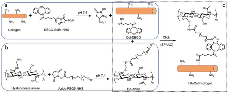
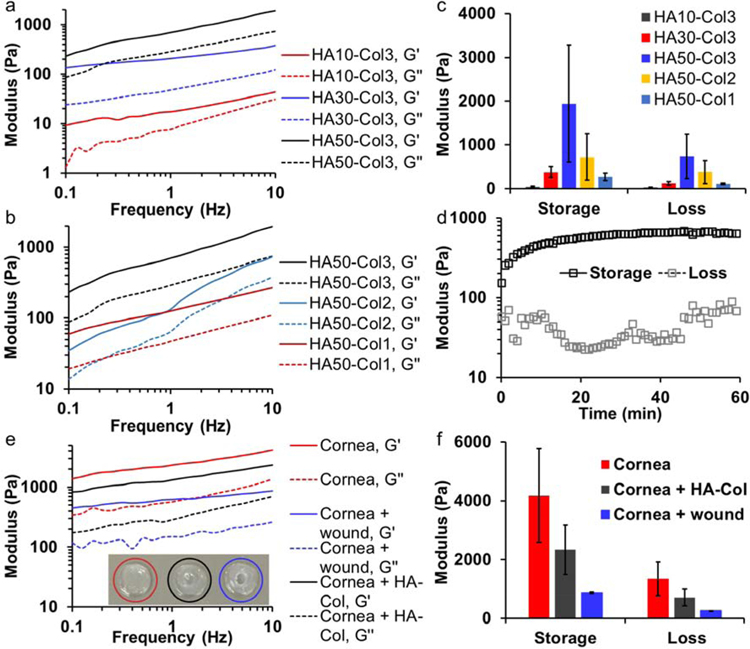
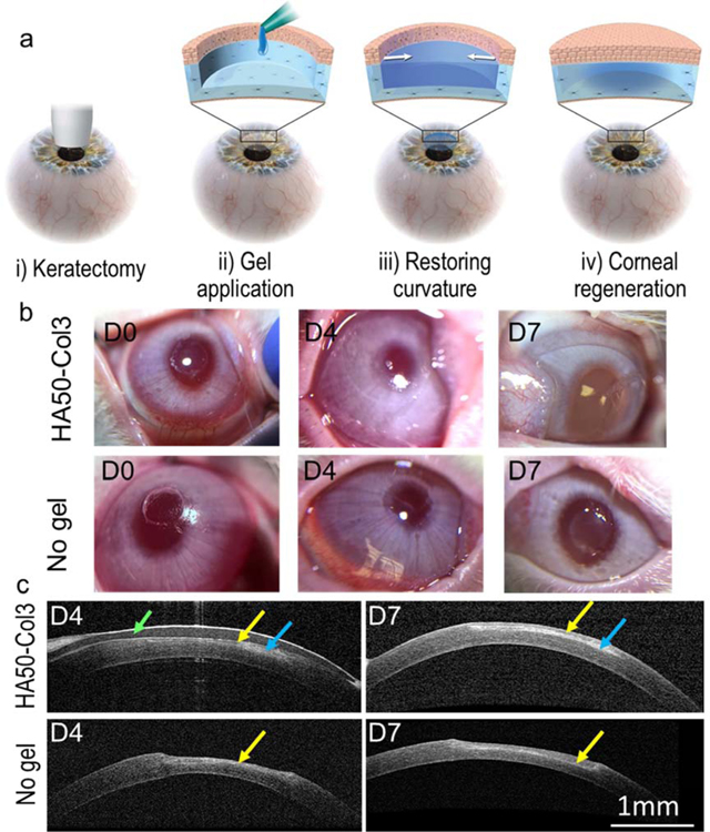
Biomaterials that mimic corneal stroma could decrease the need for donor corneal tissue and could decrease the prevalence of complications associated with corneal transplantation, including infection and rejection. We developed a bio-orthogonally crosslinked hyaluronate-collagen hydrogel which can fill corneal defects in situ without the need for any sutures, initiators, or catalysts. We studied the effects of biorthogonal crosslinking on the light transmittance of the hydrogel, which was greater than 97% water. The transmittance of the optimized hydrogel in the visible light range was over 94%. We also investigated the mechanical properties, refractive index, morphology, biocompatibility, and corneal re-epithelialization capacity of the hyaluronate-collagen hydrogel. Our in vitro, in vivo, and ex vivo results demonstrated that this bio-orthogonally crosslinked hyaluronate-collagen hydrogel has excellent potential as a biomaterial for cornea repair and regeneration.

Keywords: Bio-orthogonal; Cornea substitute; Corneal wound repair; Hyaluronate-collagen hydrogel; SPAAC crosslinking; Sutureless repair.


Conflict of interest statement

Conflict of interests

The authors Fang Chen, Gabriella Fernandes-Cunha, David Myung, and Sarah Heilshorn are co-inventors on a related patent application.

Declaration of interests

The authors declare that they have no known competing financial interests or personal relationships that could have appeared to influence the work reported in this paper.

Figures

Fig. 1
Fig. 1
Hyaluronate-collagen hydrogel crosslinked via strain-promoted azide-alkyne cycloaddition (SPAAC). (a–c) Scheme of the hydrogel formation. (a) Primary amine groups on collagen react with dibenzocyclooctyne-sulfo-N-hydroxysuccinimidyl ester to form collagen-DBCO (Col-DBCO). (b) Amidated hyaluronate react with azido-PEG5-NHS and formed hyaluronate-azide (HA-azide). (c) The SPAAC reaction between DBCO and azide groups formed hyaluronate-collagen networks without producing any byproducts.
Fig. 2
Fig. 2
Mechanical properties of hyaluronate-collagen hydrogels. (a & b) Storage and loss moduli of hydrogels increased as the frequency increased. The samples had different (a) hyaluronate (HA) and (b) collagen (Col) content, numbers are the polymer concentration (mg/mL) prior to mixing at a 1:1 volume ratio. G’ is storage modulus and G’’ is loss modulus. (c) Hydrogel with a final concentration of 25 mg/mL hyaluronate and 1.5 mg/mL collagen (HA50-Col3) showed the highest storage and loss moduli at a frequency 10 Hz. (d) Moduli of hydrogel HA50-Col3 as a function of time at the beginning of gelation. (e) Storage and loss moduli of intact and wounded cornea buttons as well as wounded cornea buttons with the stiffest hydrogel from (c). (f) The hydrogel HA50-Col3 increased the storage and loss moduli of wounded cornea buttons.
Fig. 3
Fig. 3
Optical properties of hyaluronate-collagen hydrogels. (a) Transmittance of SPAAC crosslinked hyaluronate-collagen hydrogels were much higher than non-crosslinked hydrogel (HA50&Neut. Col3). (b) Refractive index, surface focal power, and surface focal length of the hydrogels and human cornea for comparison.
Fig. 4
Fig. 4
In vitro degradation of HA50-Col3 in presence of collagenase and hyaluronidase. The HA50-Col3 degraded the fastest and most when both enzymes were present. No degradation of the gel was detected in PBS only within 24 hours.
Fig. 5
Fig. 5
Cytocompatibility, bio-integration, and re-epithelialization capacity of HA50-Col3 hydrogel to corneal epithelial cells. (a) Cell viability assay showed that collagen-DBCO 1.5 mg/mL was cytocompatible. The HA-azide at low concentrations were cytocompatible, but HA-azide at 25 mg/mL decreased the cell viability to 80%. The decrease is significant when compared to the cell only group. (* p < 0.05, *** p < 0.005, 2-tail homoscedastic t-test). The error bars represent standard deviation of four replicates. (b) In vitro live/dead assay showed that corneal epithelial cells could grow on top of the HA50-Col3 with a high survival. The hydrogel was modified with Alexa Fluor 647 (blue). Green and red showed live and dead cells, respectively. (c) Quantification of cell survival on top of HA50-Col3 hydrogel on different days based on the live/dead assay. Error bars represents standard deviations of at least five fields of views from different replicates. (d) SEM image of HA50-Col3 filling the keratectomy defect on corneal stroma. The red line roughly indicates the interface between the hydrogel and the corneal stroma. (e) Higher magnified SEM image showing the dotted area in (d). There is no detectable gap or separation between the hydrogel and the adjacent cornea, which indicates good physical apposition between the gel and the corneal stroma. (f) Top view of wounded cornea treated with and without HA50-Col3 hydrogel in an ex vivo organ culture model of sutureless anterior lamellar keratoplasty. HA50-Col promoted the re-epithelialization. The hydrogel was modified with Alexa Fluor 647 and is shown in red. Green and blue showed the F-actin and nuclei, respectively. The white arrows point to gaps in the epithelial layer on the wounded cornea without hydrogel treatment.
Fig. 6
Fig. 6
Application of hydrogel for sutureless repair of corneal wound. (a) Scheme of the disease model and treatment. The corneal wound was created by anterior partial stromal keratectomy via a customized 3.5-mm vacuum trephine. (b) Follow up photos of the rabbit eyes after in vivo keratectomy and gel application. (c) Follow-up examination of rabbit eyes with in vivo OCT. A contact lens (indicated by the green arrow) was used to protect the hydrogel (blue arrows) from scratching by the animal. The hydrogel HA50-Col3 restored the central corneal curvature. Regenerated epithelium (indicated by the yellow arrows) was seen over the keratectomy area.
Fig. 7
Fig. 7
Immunofluorescence staining of wounded cornea treated with and without hydrogel HA50-Col3. F-actin staining images of (a) wounded cornea treated with HA50-Col3, (b) wounded cornea without gel treatment, and (c) normal cornea. The results show that the HA50- Col3 effectively supports the regeneration of a thin epithelial layer. α-SMA staining images of wounded cornea treated with (d) and without (e) HA50-Col3 gel. The α-SMA staining (red) indicated that HA50-Col3 prohibited the expression of α-SMA and may prevent corneal scarring. ZO-1 staining images of (f) wounded cornea treated with HA50-Col3 gel and (g) normal cornea. The HA50-Col3 gel promoted multi-layered epithelialization with excellent tight junction ZO-1 (white) formation (f), similar to that seen in normal corneal epithelium (g).

References

    1. Islam MM, Buznyk O, Reddy JC, Pasyechnikova N, Alarcon EI, Hayes S, Lewis P, Fagerholm P, He C, Iakymenko S, Liu W, Meek KM, Sangwan VS, Griffith M, Biomaterials-enabled cornea regeneration in patients at high risk for rejection of donor tissue transplantation, npj Regenerative Medicine 3(1) (2018) 2. - PMC - PubMed
    1. Gain P, Jullienne R, He ZG, Aldossary M, Acquart S, Cognasse F, Thuret G, Global Survey of Corneal Transplantation and Eye Banking, Jama Ophthalmology 134(2) (2016) 167–173. - PubMed
    1. Dana MR, Qian Y, Hamrah P, Twenty-five–Year Panorama of Corneal Immunology: Emerging Concepts in the Immunopathogenesis of Microbial Keratitis, Peripheral Ulcerative Keratitis, and Corneal Transplant Rejection, Cornea 19(5) (2000) 625–643. - PubMed
    1. Fernandez MM, Afshari NA, Endothelial Keratoplasty: From DLEK to DMEK, Middle East Afr J Ophthalmol 17(1) (2010) 5–8. - PMC - PubMed
    1. 5–7.

Publication types