

Pursuing Experimental Reproducibility: An Efficient Protocol for the Preparation of Cerebrospinal Fluid Samples for NMR-based Metabolomics and Analysis of Sample Degradation
- PMID: 32560109
- PMCID: PMC7345835
- DOI: 10.3390/metabo10060251
Pursuing Experimental Reproducibility: An Efficient Protocol for the Preparation of Cerebrospinal Fluid Samples for NMR-based Metabolomics and Analysis of Sample Degradation
Abstract
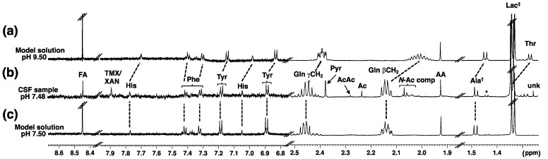
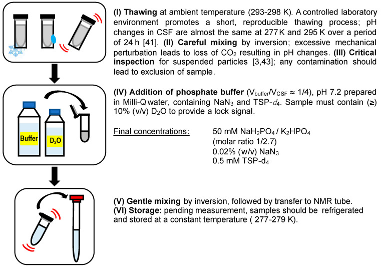
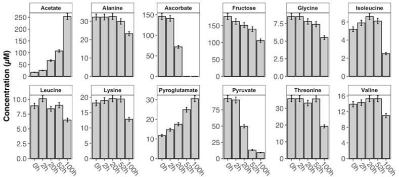
NMR-based metabolomics investigations of human biofluids offer great potential to uncover new biomarkers. In contrast to protocols for sample collection and biobanking, procedures for sample preparation prior to NMR measurements are still heterogeneous, thus compromising the comparability of the resulting data. Herein, we present results of an investigation of the handling of cerebrospinal fluid (CSF) samples for NMR metabolomics research. Origins of commonly observed problems when conducting NMR experiments on this type of sample are addressed, and suitable experimental conditions in terms of sample preparation and pH control are discussed. Sample stability was assessed by monitoring the degradation of CSF samples by NMR, hereby identifying metabolite candidates, which are potentially affected by sample storage. A protocol was devised yielding consistent spectroscopic data as well as achieving overall sample stability for robust analysis. We present easy to adopt standard operating procedures with the aim to establish a shared sample handling strategy that facilitates and promotes inter-laboratory comparison, and the analysis of sample degradation provides new insights into sample stability.
Keywords: CSF; NMR metabolomics; SOP; cerebrospinal fluid; pH; sample degradation; sample preparation; standard operating procedures; time and temperature dependence.
Conflict of interest statement
The authors declare no conflict of interest.
Figures
References
-
- Milhorat T.H., Hammock M.K. Cerebrospinal fluid as reflection of internal milieu of brain. In: Wood J.H., editor. Neurobiology of Cerebrospinal Fluid. 1st ed. Springer; Boston, MA, USA: 1983. pp. 1–23.
-
- Holmes E., Tsang T.M., Huang J.T.J., Leweke F.M., Koethe D., Gerth C.W., Nolden B.M., Gross S., Schreiber D., Nicholson J.K., et al. Metabolic profiling of CSF: Evidence that early intervention may impact on disease progression and outcome in schizophrenia. PLoS Med. 2006;3:e327. doi: 10.1371/journal.pmed.0030327. - DOI - PMC - PubMed
-
- Blasco H., Nadal-Desbarats L., Pradat P.-F., Gordon P.H., Antar C., Veyrat-Durebex C., Moreau C., Devos D., Mavel S., Emond P., et al. Untargeted 1H-NMR metabolomics in CSF: Toward a diagnostic biomarker for motor neuron disease. Neurology. 2014;82:1167–1174. doi: 10.1212/WNL.0000000000000274. - DOI - PubMed
Grants and funding
LinkOut - more resources
Full Text Sources
