. 2020 Jun 16;9(6):1475.
doi: 10.3390/cells9061475.

Inhibition of Transglutaminase 2 but Not of MDM2 Has a Significant Therapeutic Effect on Renal Cell Carcinoma

Affiliations

Inhibition of Transglutaminase 2 but Not of MDM2 Has a Significant Therapeutic Effect on Renal Cell Carcinoma

Joon Hee Kang et al. Cells. .

Abstract

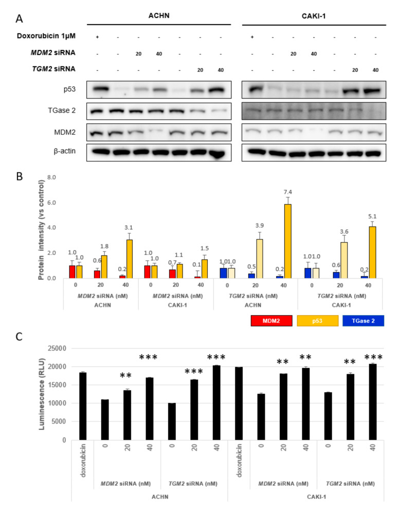
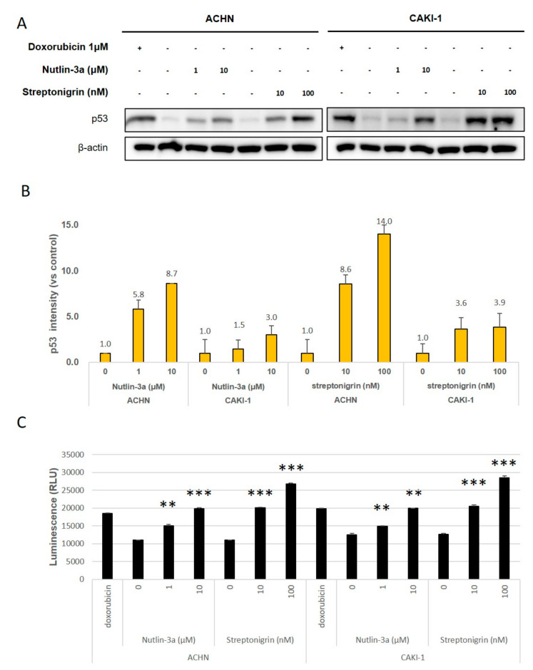
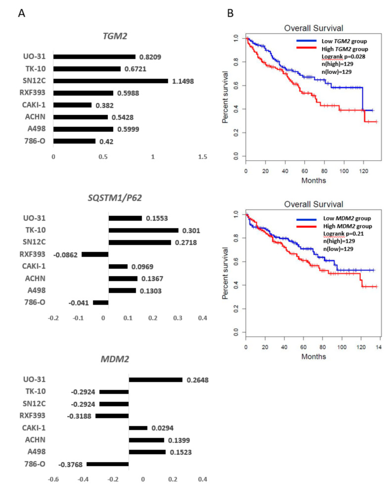
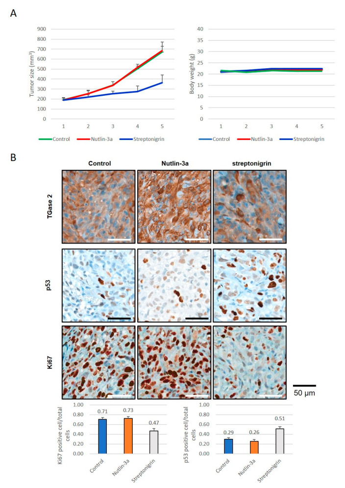
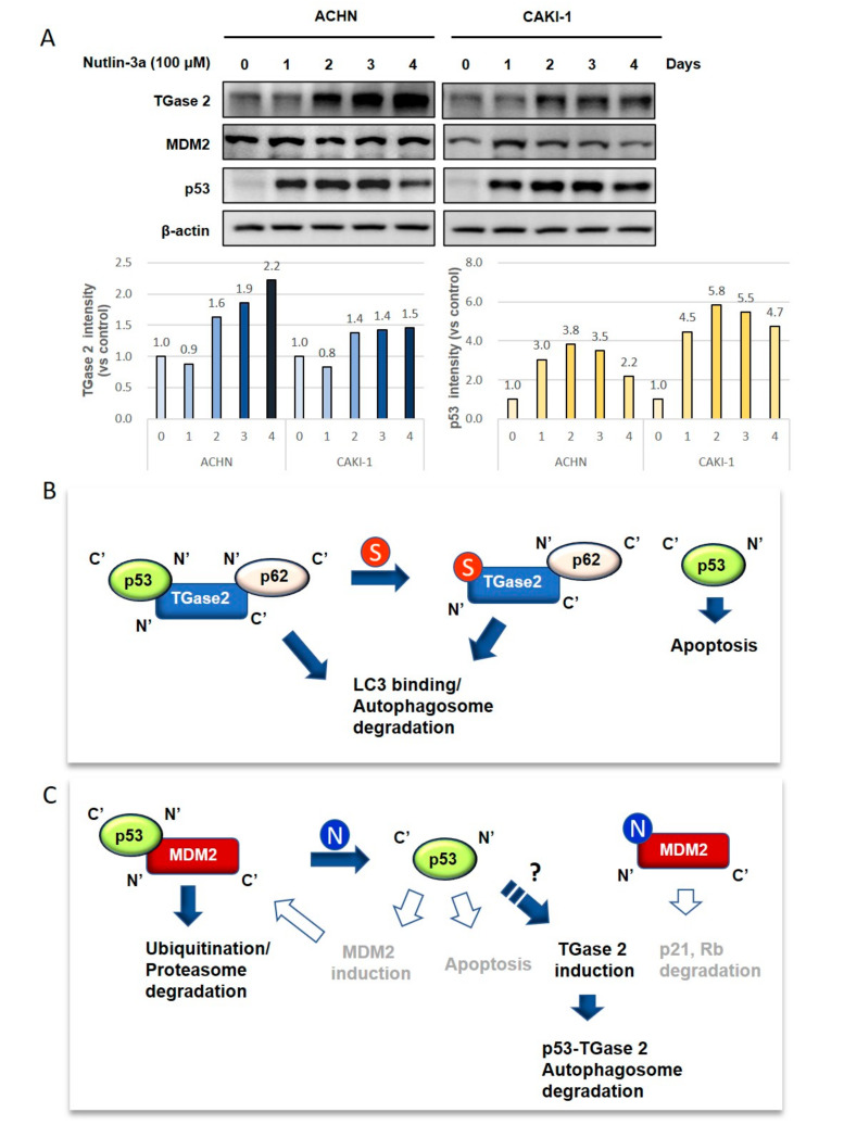
More than 50% of human cancers harbor TP53 mutations and increased expression of Mouse double minute 2 homolog(MDM2), which contribute to cancer progression and drug resistance. Renal cell carcinoma (RCC) has an unusually high incidence of wild-type p53, with a mutation rate of less than 4%. MDM2 is master regulator of apoptosis in cancer cells, which is triggered through proteasomal degradation of wild-type p53. Recently, we found that p53 protein levels in RCC are regulated by autophagic degradation. Transglutaminase 2 (TGase 2) was responsible for p53 degradation through this pathway. Knocking down TGase 2 increased p53-mediated apoptosis in RCC. Therefore, we asked whether depleting p53 from RCC cells occurs via MDM2-mediated proteasomal degradation or via TGase 2-mediated autophagic degradation. In vitro gene knockdown experiments revealed that stability of p53 in RCC was inversely related to levels of both MDM2 and TGase 2 protein. Therefore, we examined the therapeutic efficacy of inhibitors of TGase 2 and MDM2 in an in vivo model of RCC. The results showed that inhibiting TGase 2 but not MDM2 had efficient anticancer effects.

Keywords: MDM2; nutlin-3a; p53; transglutaminase 2.


Conflict of interest statement

The authors declare no conflict of interest.

Figures

Figure 1
Figure 1
Transglutaminase 2 (TGase 2) and Mouse double minute 2 homolog (MDM2) destabilize p53 in renal cell carcinoma (RCC) under normoxic conditions. (A) ACHN (left) and CAKI-1 (right) cells were transfected for 24 h with siRNA targeting TGM2 or MDM2. Doxorubicin (1 μM) was used as a positive control. (B) Image J analysis of western blots in A. (C) Apoptosis assay to assess siTGM2 or siMDM2 silencing, as monitored by RealTime-Glo™ Annexin V under the same conditions as in A. Cumulative data from three independent experiments are shown (mean ± SD; n = 3). **p < 0.01 and ***p < 0.001.
Figure 2
Figure 2
Streptonigrin and nutlin-3a increase p53 protein levels in a dose-dependent manner. (A) ACHN (left) and CAKI-1 (right) cells were treated with streptonigrin (which inhibits TGase 2-p53 binding) or nutlin-3a (which inhibits MDM2-p53 binding) for 24 h. Doxorubicin (1 μM) was used as a positive control. (B) Image J analysis of the western blots in A. (C) The apoptosis assay was performed in the presence/absence of streptonigrin or nutlin-3a and was monitored by RealTime-Glo™ annexin V under the conditions outlined in A. Cumulative data from three independent experiments are shown (mean ± SD; n = 3). **p < 0.01 and ***p < 0.001.
Figure 3
Figure 3
Expression of TGM2, MDM2, and SQSTM1/p62 in RCC: (A) Microarray data from the U.S. National Cancer Institute (http://dtp.nci.nih.gov/mtweb/targetdata). TGM2 (experiment ID: 9993; https://dtp.cancer.gov/mtweb/targetinfo?moltid=GC19395&moltnbr=9993) expression is consistently higher in RCC cell lines, whereas that of MDM2 (experiment ID: 3080; https://dtp.cancer.gov/mtweb/targetinfo?moltid=GC12482&moltnbr=3080) expression is not. Expression of SQSTM1/P62 (experiment ID: 9233; https://dtp.cancer.gov/mtweb/targetinfo?moltid=GC18635&moltnbr=9233) is higher in most RCC cell lines. The graph is on a log scale (mRNA levels in cell line/mRNA levels in a reference pool). Reference probes were made by pooling equal amounts of mRNA from the HL-60, K562, NCI-H226, COLO205, SNB-19, LOX-IMVI, OVCAR-3, OVCAR-4, CAKI-1, PC-3, MCF7, and Hs578T cell lines. The average mRNA levels of TGM2, SQSTM1/P62, and MDM2 are 0.05, −0.05, and −0.09, respectively. (B) Kaplan–Meier survival curves based on expression of TGM2 or MDM2: Patients with high expressions of TGM 2 show shorter overall survival than those with low expression (p = 0.028). The overall survival of MDM2 is not significant (p = 0.21).
Figure 4
Figure 4
The anticancer effects of inhibition of TGase 2 and MDM2 in a human RCC xenograft model: (A) ACHN cells were injected subcutaneously into one flank of BALB/c nude mice (n = 5). Mice received streptonigrin or nutlin-3a when tumors reached a volume of 200 mm3. The average tumor volume and body weight is presented as the mean ± SE. (B) ACHN cells harvested from BALB/c nude mice were analyzed by immunohistochemical staining for Ki67, TGase 2, and p53. The bar graph represents the percentage of cells positive for Ki67 or p53. Cumulative data from three independent experiments are shown (mean ± SD; n = 3). **p < 0.01 and ***p < 0.001.
Figure 5
Figure 5
The proposed model explaining p53 regulation by TGase 2 and MDM2 in RCC: (A) TGase 2 expression was induced after MDM2 inhibition. ACHN and CAKI-1 cell were treated with nutlin-3a (10 μM) in a time-dependent manner. (B) TGase 2 forms a triple complex with p53 and p62, which then moves to the autophagosome via binding of p62 to LC3. [15]. Next, p53 is degraded by the autophagic process. Streptonigrin inhibits TGase 2-p53 binding, which stabilizes p53 and triggers p53-mediated cell death. (C) P53-MDM2 binding causes proteasomal degradation of p53 via ubiquitination. Nutlin-3a inhibits the MDM2–p53 interaction, thereby inducing p53-mediated apoptosis. However, activated p53 may induce MDM2 transcription, which regulates p53 activity. This creates a feedback loop between p53 and MDM2 [28]. Therefore, MDM2 inhibition may have a temporal effect on p53 activation. The released MDM2 also triggers degradation of tumor suppressors such as p21 and Rb, which can lead to uncontrolled cell growth. In this study, we observed only increased TGase 2 expression after nultlin-3a treatment, which is likely induced by p53 activity (bold). S, streptonigrin; N, nutlin-3a; closed arrows and bold font denote observations from this study; and open arrows and gray font denote information obtained from references but failed to observe changes (data not shown). LC3, microtubule-associated proteins 1A/1B light chain 3.

References

    1. Polanski R., Noon A.P., Blaydes J., Phillips A., Rubbi C.P., Parsons K., Vlatkovic N., Boyd M.T. Senescence induction in renal carcinoma cells by Nutlin-3: a potential therapeutic strategy based on MDM2 antagonism. Cancer Lett. 2014;353:211–219. doi: 10.1016/j.canlet.2014.07.024. - DOI - PubMed
    1. Kang J.H., Lee J.S., Hong D., Lee S.H., Kim N., Lee W.K., Sung T.W., Gong Y.D., Kim S.Y. Renal cell carcinoma escapes death by p53 depletion through transglutaminase 2-chaperoned autophagy. Cell Death Dis. 2016;7:e2163. doi: 10.1038/cddis.2016.14. - DOI - PMC - PubMed
    1. Oliner J.D., Kinzler K.W., Meltzer P.S., George D.L., Vogelstein B. Amplification of a gene encoding a p53-associated protein in human sarcomas. Nature. 1992;358:80–83. doi: 10.1038/358080a0. - DOI - PubMed
    1. Iwakuma T., Lozano G. MDM2, an introduction. Mol. Cancer Res. 2003;1:993–1000. - PubMed
    1. Capoulade C., Bressac-de Paillerets B., Lefrere I., Ronsin M., Feunteun J., Tursz T., Wiels J. Overexpression of MDM2, due to enhanced translation, results in inactivation of wild-type p53 in Burkitt’s lymphoma cells. Oncogene. 1998;16:1603–1610. doi: 10.1038/sj.onc.1201702. - DOI - PubMed

Publication types

MeSH terms

Substances