Review
. 2020 Jun 17;10(6):917.
doi: 10.3390/biom10060917.

Against Repurposing Methadone for Glioblastoma Therapy

Affiliations
Review

Against Repurposing Methadone for Glioblastoma Therapy

Tatjana Vatter et al. Biomolecules. .

Abstract

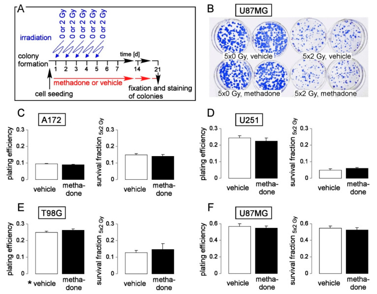
Methadone, which is used as maintenance medication for outpatient treatment of opioid dependence or as an analgesic drug, has been suggested by preclinical in vitro and mouse studies to induce cell death and sensitivity to chemo- or radiotherapy in leukemia, glioblastoma, and carcinoma cells. These data together with episodical public reports on long-term surviving cancer patients who use methadone led to a hype of methadone as an anti-cancer drug in social and public media. However, clinical evidence for a tumoricidal effect of methadone is missing and prospective clinical trials, except in colorectal cancer, are not envisaged because of the limited preclinical data available. The present article reviews the pharmacokinetics, potential molecular targets, as well as the evidence for a tumoricidal effect of methadone in view of the therapeutically achievable doses in the brain. Moreover, it provides original in vitro data showing that methadone at clinically relevant concentrations fails to impair clonogenicity or radioresistance of glioblastoma cells.

Keywords: A172; T98G; U251; U87MG; cell cycle regulation; clonogenic survival; colony formation assay; flow cytometry; human glioblastoma cell lines; ionizing radiation.


Conflict of interest statement

D.Z. and F.E. have research and educational grants from Elekta, Philips, Siemens, Sennewald. The other authors declare no competing interests.

Figures

Figure 1
Figure 1
Expression of methadone target molecules in glioblastoma (Illumina HiSeq_RNA Seq V2 glioblastoma dataset (n = 172) of The Cancer Genome Atlas (TCGA)). (A) Majority of glioblastoma specimens exhibit non-detectable low abundances of the µ-opioid receptor (OPRM1) mRNA. Here, the µ-opioid receptor mRNA abundances are plotted against those of the GALR1 (galanin receptor 1), which forms heteromeric receptors with OPRM1 (values of individual tumors are shown). (B) Box whisker plots depicting relative mRNA abundances (TCGA-normalized values) of proposed methadone targets in glioblastoma specimens (medians are highlighted by red lines). Cav channels: voltage-gated L- and T-type Ca2+ channels, nAChRs: nicotinic acetylcholine receptors, NMDA receptors: N-methyl-d-aspartate receptors, Nav channels: voltage-gated Na+ channels, KCNH2: hERG1 K+ channel, CLCN2: ClC-2 Cl channel, ABCB1: p-glycoprotein (MDR1).
Figure 2
Figure 2
Abundance of µ-opioid (OPRM1) and N-methyl-d-aspartate (NMDA) receptors (GRIN1) in human glioblastoma cell lines. (A) Mean (SE, n = 3) housekeeper-normalized abundances of OPRM1 (left) and GRIN1 (right) mRNA in A172, T98G, and U251 cells. (B) Immunoblots from total cell lysates of U251 (left) and T98G (right) cells separated by SDS-PAGE and probed against OPRM1 (μ-opioid receptor, upper blot) and for loading control against β-actin (lower blot) Human glioblastoma cell lines T98G, A172 and U87MG (see below) were obtained from the American Type Culture Collection (ATCC, Bethesda, MD, USA) and grown in 10% fetal bovine serum (FBS)-supplemented RPMI-1640 medium. The U251 cells were a kind gift from Dr. Luiz O. Penalva (Graduate School of Biomedical Sciences, UT Health San Antonio, Texas) and grown in Dulbecco’s modified Eagle’s medium containing 4500 mg glucose/l and 10% FBS. Total RNA was isolated using the NucleoSpin RNA Kit (Macherey-Nagel, Düren, Germany). OPRM1-, GRIN1- and housekeeper β-actin (ACTB)-, pyruvate dehydrogenase beta (PDHB)-, and glyceraldehyde-3-phosphate dehydrogenase (GAPDH)-specific fragments were amplified by the use of SYBR Green-based quantitative real-time PCR (QuantiTect Primer Assay QT00001512, QT00082089, QT00095431, QT00031227, and QT01192646, QIAGEN, Venlo, Netherlands, and 1Step RT qPCR Green ROX L Kit, highQu, Kraichtal, Germany) in a Roche LightCycler® 480 (Roche, Mannheim, Germany). Abundances of the individual mRNAs were normalized to the geometrical mean of the three housekeeper mRNAs. For western blotting, cells were lysed in a buffer (containing in mM: 50 N-2-hydroxyethylpiperazine-N-2-ethanesulfonic acid (HEPES) pH 7.5, 150 NaCl, 1 ethylenediaminetetraacetic acid (EDTA), 10 sodium pyrophosphate, 10 NaF, 2 Na3VO4, 1 phenylmethylsulfonylfluoride (PMSF) additionally containing 1% triton X-100, 5 µg/mL aprotinin, 5 µg/mL leupeptin, and 3 µg/mL pepstatin) and separated by SDS-PAGE under reducing condition. Blots were blocked in tris(hydroxymethyl)aminomethane-buffered saline (TBS) containing 0.05% Tween 20 and 5% non-fat dry milk for 1 h at room temperature. The membrane was incubated overnight at 4 °C and for 1 h at room temperature with recombinant anti-µ-opioid receptor antibody [UMB3] [96] (ab227067, Abcam, Berlin, Germany, 1:500) and anti-β-actin (1:20,000, clone AC-74, #A2228, Sigma-Aldrich, Deisenhofen, Germany), respectively. Antibody binding was detected with a horseradish peroxidase-linked goat anti-rabbit IgG antibody or anti-mouse IgG antibody (Cell Signaling # 7074 and # 7076, respectively; 1:1000–1:2000) incubated for 1 h at room temperature (all antibody dilutions in TBS-Tween/5% milk) and enhanced chemiluminescence (ECL western blotting analysis system, GE Healthcare/Amersham-Biosciences, Freiburg, Germany).
Figure 3
Figure 3
High-dose methadone (20 µM) may modify cell cycle progression in human glioblastoma cells. (A) Histograms of permeabilized, propidium iodide-stained T98G glioblastoma cells recorded by flow cytometry (Nicoletti protocol) 24 h after irradiation with 0 Gy (left) or 4 Gy (right). Cells were pre- (1 h) and co-incubated (24 h) either with 0 μM (vehicle, black) or 20 μM (red) methadone. (BD) Mean (± SE, n = 6) percentage of A172 (B), T98G (C) and U251 (D) glioblastoma cells that reside in G1 (upper row), S (middle row) and G2 (lower row) phase of cell cycle 24 h (left) and 48 h (right) after irradiation with 0 or 4 Gy. Cells were pre-incubated (1 h), irradiated and post-incubated (24 or 48 h) in the presence of 0 μM (vehicle, open bars) or 20 μM (closed bars) methadone. * indicates zp ≤ 0.05, two-tailed (Welch-corrected) t-test with Bonferroni correction for z = 16 pairwise comparisons. Cells were irradiated (6 MV photons, single dose of 0 or 4 Gy) using a linear accelerator (LINAC SL25 Philips) at a dose rate of 4 Gy/min at room temperature and post-incubated for further 24 h and 48 h in the absence (vehicle, ethanol) or presence of methadone (20 µM). For cell cycle analysis, cells were permeabilized and stained (30 min at room temperature) with propidium iodide solution (containing 0.1% Na-citrate, 0.1% triton X-100, 10 µg/mL propidium iodide in phosphate-buffered saline, PBS), and the DNA amount was analyzed by flow cytometry (FACS Calibur, Becton Dickinson, Heidelberg, Germany, 488 nm excitation wavelength) in fluorescence channel FL-3 (linear scale, >670 nm emission wavelength). Data were analyzed with the FCS Express 3 software (De Novo Software, Los Angeles, CA, USA).
Figure 4
Figure 4
Heterogeneous effects of high-dose methadone (20 µM) on clonogenic survival of irradiated (0–8 Gy) human A172 (AD), T98G (EH) an U251 (IL) glioblastoma cells. (A,E,I) Coomassie blue-stained colonies regrown from single 0 Gy (upper row)- and 6 Gy (lower row)-irradiated A172 (A), T98G (E), and U251 (I) glioblastoma cells pre- (1 h) and post-incubated (24 h) with either 0 μM (vehicle, ethanol, left) or 20 μM (right) methadone. (B, F, J) Mean (± SE, n = 11–12) plating efficiency of A172 (B), T98G (F), and U251 (J) glioblastoma cells incubated (24 h) with either 0 μM (vehicle, open bar) or 20 μM (closed bar) methadone. (C,D,G,H,K,L) Mean (± SE, n = 11–12) survival fraction of irradiated (0, 2, 4, 6, and 8 Gy in (C,G,K) and 2 Gy in (D,H,L)) A172 (C,D), T98G (G,H), and U251 (K,L) cells irradiated and pre- (1 h) as well as post-incubated (24 h) with either 0 μM (vehicle, open circles and open bars) or 20 μM (red triangles and closed bars) methadone. * indicates p ≤ 0.05, two-tailed (Welch-corrected) t-test. For delayed plating colony formation, cells were irradiated (0, 2, 4, 6, 8 Gy) and post-incubated for 24 h in the absence (vehicle) or presence of methadone (20 µM). Then, cells were replated in methadone-free medium and grown for a further two weeks. Thereafter, colonies were defined as cell clusters of ≥50 cells and colony numbers were counted. Plating efficiency (PE) was defined by the ratio between counted colonies (N) and plated cells (n) under control conditions (PE = N/n). The survival fraction (SF) was calculated by normalizing in both arms (methadone and vehicle control) separately the plating efficiency after irradiation (PExGy) to that of the corresponding unirradiated control (PE0Gy) by the formula SF = PExGy/PE0Gy. SFs were fitted by the use of the linear quadratic equation (SF = e-(α·D + β·D^2) with D = radiation dose and α and β cell-specific constants).
Figure 5
Figure 5
Methadone in a clinically relevant concentration (2 µM) does not impair clonogenic survival or radioresistance of human glioblastoma cells. (A) Experimental protocol of pre-plating colony formation depending on methadone concentration (0 or 2 µM) and fractionated irradiation (5 × 0 or 5 × 2 Gy). (B) Coomassie blue-stained colonies regrown from 5 × 0 Gy (left)- and 5 × 2 Gy (right) U87MG cells co-incubated with either 0 μM (vehicle, ethanol, upper row) or 2 μM (lower row) methadone. (CF). Mean (± SE, n = 6) plating efficiency (left) and survival fraction 5 × 2 Gy (right) of A172 (C), T98G (D), U251 (E), and U87MG (F) glioblastoma cells co-incubated with either 0 μM (vehicle, open bars) or 2 μM methadone (closed bars). For pre-plating colony formation, plated cells were irradiated (0 or 2 Gy on days 1, 2, 3, 4, and 5) and post-incubated in the absence (vehicle, ethanol) or presence of methadone (2 µM) until formation of colonies. Thereafter, plating efficiencies and survival fractions were calculated as described in the legend of Figure 4.

References

    1. Wolff P. On pethidine and methadone derivatives. Bull. World Health Organ. 1949;2:193. - PMC - PubMed
    1. Bockmühl M., Ehrhart G. Über eine neue Klasse von spasmolytisch und analgetisch wirkenden Verbindungen, I. Justus Liebigs Ann. Chem. 1949;561:52–85. doi: 10.1002/jlac.19495610107. - DOI
    1. Scott C.C., Chen K. The action of 1, 1-diphenyl-1-(dimethylaminoisopropyl)-butanone-2, a potent analgesic agent. J. Pharmacol. Exp. Ther. 1946;87:63–71. doi: 10.1097/00000542-194609000-00028. - DOI - PubMed
    1. Kreek M.J., Vocci F.J. History and current status of opioid maintenance treatments: Blending conference session. J. Subst. Abuse Treat. 2002;23:93–105. doi: 10.1016/S0740-5472(02)00259-3. - DOI - PubMed
    1. Dole V.P., Nyswander M. A medical treatment for diacetylmorphine (heroin) addiction. A clinical trial with methadone hydrochloride. JAMA. 1965;193:646–650. doi: 10.1001/jama.1965.03090080008002. - DOI - PubMed

Publication types

MeSH terms