Review
. 2020 Jun 17;21(12):4320.
doi: 10.3390/ijms21124320.

Telocytes in the Normal and Pathological Peripheral Nervous System

Affiliations
Review

Telocytes in the Normal and Pathological Peripheral Nervous System

Lucio Díaz-Flores et al. Int J Mol Sci. .

Abstract

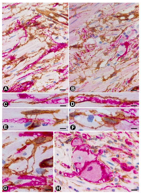
We studied telocytes/CD34+ stromal cells in the normal and pathological peripheral nervous system (PNS), for which we reviewed the literature and contributed our observations under light and electron microscopy in this field. We consider the following aspects: (A) general characteristics of telocytes and the terminology used for these cells (e.g., endoneurial stromal cells) in PNS; (B) the presence, characteristics and arrangement of telocytes in the normal PNS, including (i) nerve epi-perineurium and endoneurium (e.g., telopodes extending into the endoneurial space); (ii) sensory nerve endings (e.g., Meissner and Pacinian corpuscles, and neuromuscular spindles); (iii) ganglia; and (iv) the intestinal autonomic nervous system; (C) the telocytes in the pathologic PNS, encompassing (i) hyperplastic neurogenic processes (neurogenic hyperplasia of the appendix and gallbladder), highly demonstrative of telocyte characteristics and relations, (ii) PNS tumours, such as neurofibroma, schwannoma, granular cell tumour and nerve sheath myxoma, and interstitial cell of Cajal-related gastrointestinal stromal tumour (GIST), (iii) tumour-invaded nerves and (iv) traumatic, metabolic, degenerative or genetic neuropathies, in which there are fewer studies on telocytes, e.g., neuroinflammation and nerves in undescended testicles (cryptorchidism), Klinefelter syndrome, crush injury, mucopolysaccharidosis II (Hunter's syndrome) and Charcot-Marie-Tooth disease.

Keywords: Meissner corpuscles; appendicular neurogenic hyperplasia; gallbladder neurogenic hyperplasia; nerves; peripheral nervous system tumours; telocytes.


Conflict of interest statement

The authors declare no conflict of interest.

Figures

Figure 1
Figure 1
In peripheral nerves, telocytes (TCs)/CD34+SCs are observed in the epi-perineurium and endoneurium under light (A,B) and electron microscopy (CF). It should be noted that in B, the TCs (brown) are arranged, underlying the perineurial cell layer (perineurial cells: red) in the epi-perineurium. C–F: Ultrastructural characteristics of endoneurial TCs, in which long, thin telopodes (C and D, arrows) and homocellular junctions (E and F, arrows) are seen. A and B: sections immunostained with anti-CD34 (A), and double-immunostained with anti-CD34 (brown) and epithelial membrane antigen (EMA) (red) (haematoxylin-stained nuclei). C–F: Ultrathin sections. Uranyl acetate and lead citrate. Bar: A, 60 µm; B, 40 µm; C, 2 µm; D, E, F, 1 µm.
Figure 2
Figure 2
Examples of TCs in some sensory receptors, ganglia and the autonomic nervous system in the digestive tract (AC). In Meissner corpuscles, TCs are observed in the capsule (A and C), showing CD34 expression (A, brown) and the characteristic ultrastructure (C, arrows). Schwann cells, with S100 expression (B, brown), are observed. It should be noted that in C, a nerve fibre (asterisk) surrounded by Schwann cells can be observed. (D,E) In neuromuscular spindles, TC telopodes (arrows) are shown around striated muscle cells. (F) A TC telopode (arrow) between a neuronal–glial unit (Neuron: N and satellite glial cell: SC) and nerve fibres in a spinal ganglion. (G) TCs expressing CD34 (brown) in an appendiceal myenteric ganglion. A, G: Sections stained with anti-CD34. B: Section stained with anti-S100. C–F. Ultrathin sections. Uranyl acetate and lead citrate. Bar: A, B, 60 µm; C, 2 µm; D, E, F, 1 µm; G, 40 µm
Figure 3
Figure 3
TCs/CD34+SCs (brown) around appendiceal hyperplastic nerve fibres (Schwann cells and axons) (red) (AG) and neuronal–glial units (B,H). Sections stained with anti-CD34 (brown) and anti-S-100 (red) (A, C–E and G and H) and anti-CD34 and anti-neurofilaments (B and D). Numerous fusiform or stellate TCs and their telopodes (brown) are observed around the aforementioned structures. The TCs and telopodes follow the path of the nerve fibres and establish contact with TCs of other nerve fibres and smooth muscle cells. It should be noted how thinner telopodes can originate from the somatic region of the TCs (Figure 3E) or from thicker and initial telopodes (Figure 3G). Bar: A, B, 20 µm; C-H, 10 µm
Figure 4
Figure 4
TCs/CD34+SCs around nerve fibres (Schwann cells and axons) and neuronal–glial units growing in the appendiceal adipose and connective tissues, and in the adventitia of blood vessels in the presence of mast cells. Sections double-immunostained with anti-CD34 (brown) and anti-S100 (red) (A), anti-CD34 (brown) and anti-neurofilaments (red) (B) and anti-CD34 (brown) and c-kit (red) (insert in B). A: TCs/CD34+SCs (brown) are observed around neuronal–glial units between adipocytes (ad). It should be noted that the satellite glial cells are stained in red. B: Nerve fibres between the TCs/CD34+SCs of blood vessel adventitia (L: vessel lumen). Insert of B: A c-kit immunostained mast cell (red) associated with a TC/CD34+SC) (brown). Bar: A, 20 µm; B, 40 µm; insert of B, 10 µm.
Figure 5
Figure 5
TCs in the neurogenic hyperplasia of gallbladder. (A) Presence of TCs/CD34+SCs (brown) around and within a thick nerve in the gallbladder wall. Schwann cells expressing S-100 (red) are seen. (B,C) TCs/CD34+SCs (brown) between fascicles and independent nerve fibres arranged in different directions within the nerves. (D) Nerve fibres (expressing neurofilaments, red) between TCs/CD34+SCs (brown) in the blood vessel adventitia. (E) Nerve fibres in the chorion of the mucosa. The absence of TCs/CD34+SCs is noted. A and E: Sections double-immunostained with anti-CD34 (brown) and anti-S100 (red). B and C: Sections immunostained with anti-CD34 (brown). D: Section double-immunostained with anti-CD34 (brown) and anti-neurofilaments (red). Bar: A, C, 80 µm; B, 100 µm; D, 20 µm; F, 30 µm.
Figure 6
Figure 6
TCs/CD34+SCs in neurofibromas. (AC) Using double-immunostaining (anti-CD34 and anti-S100), numerous TCs/CD34+SCs (brown), intermixed with Schwann cells (red) are observed forming bundles with parallel, arciform or irregular arrangement. (D) TCs/CD34+SCs are also seen in the adventitia of blood vessels in the tumour. (E) CD34+ cells (ameboid dendritic cells) in a myxoid area of a plexiform neurofibroma. D and E: Immunostained with anti-CD34. Bar: A, 80 µm; B, D, 60 µm; C, 20 µm; E, 40 µm.
Figure 7
Figure 7
Multi-vacuolated cells (ameboid dendritic cells) in myxoid areas of plexiform neurofibromas. (AE) CD34 expression in the vacuolated cells, which partially retain their primitive fusiform or stellate morphology (A–C) or acquire a globe-like aspect, sometimes with a piriform appearance (D and E). (F) Alcian blue positivity in the extracellular matrix and in the vacuolated cells (G,H) Ultrastructural characteristics of the cells in the myxoid areas. Note the intracytoplasmic vacuoles and how one cell retains some processes (G), while the other acquires a globoid-like aspect (H). A–E: Anti-CD34 immunostaining. F: Alcian blue staining. G and H: Ultrathin sections. Uranyl acetate and lead citrate. Bar: A–E, 5 µm; F, 10 µm; G, H, 0.5 µm.
Figure 8
Figure 8
TCs/CD34+SCs in schwannomas and granular cell tumours. (AC) A schwannoma in which numerous TCs/CD34+SCs (brown) (A and B) are arranged around groups of Schwann cells, which form Verocay bodies (A and B) and express S100 (brown) (C). D and E: Granular cell tumour, in which TCs/CD34+SCs) (brown) (D) surround granular cells (granular S100-positive Schwann cells) (brown) (E). (F,G) Ultrastructural characteristics of the granular cells in whose environment some telopodes are observed (arrows). A to E: Sections immunostained with anti-CD34 (brown) (A, B, D) and anti S-100 (brown) (C and E). F and G: Ultrathin sections, Uranyl Acetate and Lead citrate. Bar: A, B, C, E, 80 µm; D, 60 µm; F, G, 0.5 µm.
Figure 9
Figure 9
TCs/CD34+SCs and S-100+ cells in nerve myxomas (AE), and CD34 and c-kit (CD-117) expression in gastrointestinal stromal tumours (GISTs) (F,G). A and B: Myxoid lobules in a nerve sheath myxoma, in which long, thin telopodes of spindled TCs/CD34+SCs are predominantly arranged in their periphery (A) surrounding S100+ Schwann cells (B). (C–E) Increased number of Meissner corpuscles in the papillary dermis above the tumour (C, expressing CD34, and D, expressing S-100) and lobules of the lesion near the epidermis, showing a morphology reminiscent of Meissner corpuscles. (E). F and G: Expression of c-kit (F) and CD34 (G) in cells of a GIST. A, C and G: Anti-CD34 immunostaining. B, D and E: Anti-S100 immunostaining. F: c-kit immunostaining. Bar: A, B, E–G, 100 µm; C, D, 60 µm.
Figure 10
Figure 10
TCs/CD34+SCs in tumour-invaded and injured (compressed) nerves, and in nerves of the testicle affected by Klinefelter syndrome. (A) An adenocarcinoma-invaded nerve (arrow = neoplastic gland) in the gallbladder with a marked increase in the number of TCs/CD34+SCs (brown). (B) A nerve (arrow) next to a neoplastic gland (g) also shows hyperplastic TCs/CD34+SCs. (C) Telopodes of a TC (arrows) in a compressed nerve. (DF) Ultrastructural characteristics of TCs (arrows) around nerve fibres in the testicles of a patient with Klinefelter syndrome. A and B: CD34 Immunostaining. C–F: Ultrathin sections. Uranyl acetate and lead citrate. Bar: A, B, 80 µm; C, E, F, 1 µm; D, 2 µm.

Similar articles

Cited by

References

    1. Popescu L.M., Faussone-Pellegrini M.S. Telocytes—A case of serendipity: The winding way from Interstitial Cells of Cajal (ICC), via Interstitial Cajal-Like Cells (ICLC) to telocytes. J. Cell. Mol. Med. 2010;14:729–740. doi: 10.1111/j.1582-4934.2010.01059.x. - DOI - PMC - PubMed
    1. Faussone-Pellegrini M.S., Popescu L.M. Telocytes. BioMol. Concepts. 2011;2:481–489. doi: 10.1515/BMC.2011.039. - DOI - PubMed
    1. Popescu L.M. Telocytes—A novel type of interstitial cells. In: Braisant O., Wakamatsu H., Kang I., Allegaert K., Lenbury Y., Wacholtz A., editors. Recent Researches in Modern Medicine—HISTEM. WSEAS Press; Cambridge, UK: 2011. pp. 424–432.
    1. Cretoiu D., Radu B.M., Banciu A., Banciu D.D., Cretoiu S.M. Telocytes heterogeneity: From cellular morphology to functional evidence. Semin. Cell Dev. Biol. 2017;64:26–39. doi: 10.1016/j.semcdb.2016.08.023. - DOI - PubMed
    1. Vannucchi M.G., Faussone-Pellegrini M.S. The telocyte subtypes. Adv. Exp. Med. Biol. 2016;913:115–126. - PubMed

MeSH terms