Review
. 2020:105:293-380.
doi: 10.1016/bs.adgen.2020.01.002. Epub 2020 May 4.

Application of yeast to studying amyloid and prion diseases

Affiliations
Review

Application of yeast to studying amyloid and prion diseases

Yury O Chernoff et al. Adv Genet. 2020.

Abstract

Amyloids are fibrous cross-β protein aggregates that are capable of proliferation via nucleated polymerization. Amyloid conformation likely represents an ancient protein fold and is linked to various biological or pathological manifestations. Self-perpetuating amyloid-based protein conformers provide a molecular basis for transmissible (infectious or heritable) protein isoforms, termed prions. Amyloids and prions, as well as other types of misfolded aggregated proteins are associated with a variety of devastating mammalian and human diseases, such as Alzheimer's, Parkinson's and Huntington's diseases, transmissible spongiform encephalopathies (TSEs), amyotrophic lateral sclerosis (ALS) and transthyretinopathies. In yeast and fungi, amyloid-based prions control phenotypically detectable heritable traits. Simplicity of cultivation requirements and availability of powerful genetic approaches makes yeast Saccharomyces cerevisiae an excellent model system for studying molecular and cellular mechanisms governing amyloid formation and propagation. Genetic techniques allowing for the expression of mammalian or human amyloidogenic and prionogenic proteins in yeast enable researchers to capitalize on yeast advantages for characterization of the properties of disease-related proteins. Chimeric constructs employing mammalian and human aggregation-prone proteins or domains, fused to fluorophores or to endogenous yeast proteins allow for cytological or phenotypic detection of disease-related protein aggregation in yeast cells. Yeast systems are amenable to high-throughput screening for antagonists of amyloid formation, propagation and/or toxicity. This review summarizes up to date achievements of yeast assays in application to studying mammalian and human disease-related aggregating proteins, and discusses both limitations and further perspectives of yeast-based strategies.

Keywords: Alzheimer's disease; Amyloid β; Amyotrophic lateral sclerosis; Huntington's disease; Parkinson's disease; Prion protein; Protein aggregation; Tau; Transthyretin; α-Synuclein.


Figures

Fig. 1
Fig. 1
Templated nucleated polymerization of amyloids and prions. The example of parallel in-register cross-β amyloid structure (β-arch) is shown. Boxes with arrowheads correspond to β-strands. The folded intermolecular β-sheet exists only within a polymer. A newly immobilized monomer acquires exact same conformation as a pre-existing unit of the amyloid fibril due to formation of hydrogen bonds between identical amino acid (aa) residues.
Fig. 2
Fig. 2
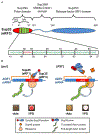
The Sup35/[PSI+] system in yeast. (A) Structural and functional organization of the yeast Sup35 protein. NQ—asparagine- and glutamine-rich stretch, NR—region of oligopeptide repeats. Repeats are indicated by green boxes. Sequences of oligopeptide repeats are shown by green characters, the piece of the sequence located between the first and second repeats—by red characters. Numbers correspond to aa positions. (B) The phenotypic detection assay for the [PSI+] prion. On the left—soluble Sup35 (eRF3), together with Sup45 (eRF1), is functioning as a part of translation termination complex in the [psi] strain bearing the premature stop codon in the ADE1 gene (UGA nonsense-allele ade1-14). Termination on this premature stop codon results in the formation of truncated Ade1 protein, leading to the inability to grow on the medium lacking adenine (−Ade) and red color (due to accumulation of the red pigment, which is a polymerized intermediate of the adenine biosynthetic pathway) on the complete organic (YPD) medium. On the right—aggregation of Sup35 in the prion-containing ([PSI+]) cells, accompanied by sequestration of Sup45, decreases the ability of the termination complex to access translating ribosomes, and results in the impairment of termination, leading to the readthrough (nonsense-suppression) of premature UGA codon and synthesis of full-length Ade1 protein, that confers growth on −Ade medium and prevents accumulation of the red pigment on YPD medium. Designations of the soluble and aggregated (prion) forms of Sup35, as well as designations of Sup45, ribosome and newly synthesized Ade1 polypeptide are indicated.
Fig. 3
Fig. 3
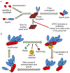
[PSI+] formation and propagation in yeast, and roles of other proteins. (A) Induction of [PSI+] formation by overproduction of constructs bearing Sup35 PrD (Sup35N or NM), in the presence of another yeast prion (such as Rnq1 prion, [PIN+]) acting as a heterologous nucleation center. (B) Chaperone role in [PSI+] propagation: fragmentation of amyloid fibrils, generating new oligomeric “seeds” for new rounds of polymerization is achieved by the chaperone machinery composed of the Hsp104, Hsp70-Ssa and Hsp40 proteins. Designations of the prion and non-prion isoforms are the same as in Fig. 2. See more detailed comments in the text.
Fig. 4
Fig. 4
Huntingtin protein (Htt) and its derivatives used to model Huntington’s disease (HD) in yeast. (A) Human full-length wild-type (WT) and Huntington disease-associated (HD) variants of the Htt protein. Aa numbering is shown for the variant with 23 glutamines (Qs) in the polyglutamine (PolyQ) tract. N17—amino terminal region of 17 aa; P-rich—proline-rich region. (B) Yeast constructs for studying polyQ aggregation and toxicity. For the polyQ-expanded version, only a construct with the longest polyQ stretch (designated as 103Q, see explanation in the text) is shown as an example. Yeast constructs with shorter polyQ expansions are also used as described in the text. GFP—green fluorescent protein (the most frequently used fluorophore, although other fluorophores are also occasionally employed). This should be noted that majority of the yeast Htt-derived polyQ constructs also contain the FLAG epitope attached at the N-terminus (not shown in figure).
Fig. 5
Fig. 5
Role of endogenous yeast prions in polyQ aggregation and cytotoxicity. (A) Rnq1 aggregates nucleate formation of multiple peripheral 103Q-GFP aggregates in the [PIN+] strain, containing Rnq1 protein in a prion form. Sequestration of the endocytosis-associated (EA) proteins by 103Q-GFP aggregates leads to the defect of endocytosis, resulting in cytotoxicity. Other proteins, sequestered by polyglutamine aggregates and possibly contributing to cytotoxicity are discussed in the text. (B) 103QP-GFP protein, containing the P-rich region (see Fig. 4), is assembled into a cytoprotective aggregate deposit (aggresome), colocalized with a spindle body. This prevents sequestration of EA proteins and makes constructs non-toxic to [psi] cells, containing Sup35 protein in a non-prion form. However, in the [PSI+] cells, which contain the prion form of Sup35 protein, 103QP-GFP polymers sequester aggregated Sup35 (and through it, another translation termination factor, Sup45), leading to the defect of translation, that results in cytotoxicity. Designations unique for this figure are shown in the bottom left corner;other designations are the same as on Fig. 2.
Fig. 6
Fig. 6
Aβ and tau proteins. (A) Generation of Aβ by proteolytic processing of the amyloid precursor protein (APP). APP processing is catalyzed by the membrane-associated secretase complex. Cleavage by β-secretase and subsequently, by γ-secretase produces Aβ peptides, while cleavage by α-secretase prevents Aβ formation. Depending of the position of β-secretase cleavage site, Aβ peptides of various lengths are produced. Two major sites, leading to the formation of 40 aa (Aβ40) and 42 aa (Aβ42) peptides are indicated. An example of Aβ42 production as well as its subsequent polymerization are shown. (B) Structural and functional organization of the longest isoform of human tau protein of 441 aa in length (tau441 or tau 2N4R) presented in the neurons of central nervous system is shown. Alternative splicing may eliminate some or all of the regions shown in blue rectangles, resulting in the generation of total of six tau isoforms, denoted by either their total number of amino acids or the number of N-terminal exons (N) and microtubule-associated repeats (R). Exons absent in some of the shorter isoforms but present in the longest isoform (N1 and N2) are termed “Inserts.” The N-terminal part tau is referred to as the “Projection domain” since it projects away from the microtubule surface and can interact with membrane-associated structures or motor proteins. Microtubule-assembly domain containing repeat sequences (R1-R4 in the longest isoform), and adjacent proline-rich (P-rich) region are also indicated. These regions of tau regulate the rate of microtubule polymerization. Repeat sequences are also involved in the formation of tau amyloid fibrils.
Fig. 7
Fig. 7
Examples of yeast model systems for the detection of Aβ aggregation. (A) Fusion of Aβ to a fluorophore: C-terminal fusions of Aβ40 and Aβ42 to green (GFP), yellow (YFP) or cyan (CFP) fluorescent proteins are shown. (B) Chimeric construct for the phenotypic detection of Aβ aggregation, using the yeast Sup35 protein (translation termination factor) as a reporter in a termination readthrough (nonsense-suppression) assay described on Fig. 2. In this construct, Aβ42 is substituted for the PrD region of Sup35 (Sup35N). Resulting chimeric protein, retaining the middle (Sup35M) and the C-proximal release factor (RF, Sup35) domains of Sup35 is termed Aβ42-MRF. Designations of the Sup35 domains are the same as on Fig. 2; Aβ designations are the same as on Fig. 6. Numbers indicate amino acid positions in Aβ (black font, located under the drawing in a chimeric construct) and Sup35 (red font, located above the drawing in a chimeric construct).
Fig. 8
Fig. 8
Mouse prion protein (PrP) and its derivatives used in yeast studies. (A) Structural and functional organization of mouse PrP(moPrP). Signal peptide (first 22 aa), which is cleaved during processing, as well as the region including G-rich octapeptide repeats (indicated by navy blue boxes), and a GPI anchoring signal (GPI) are indicated. The processed form of moPrP (23–230) includes the N-proximal unstructured domain and C-terminal globular domain as shown. The globular domain contains three α-helices (α1–3), and two β-strands (β1–2) as indicated. Glycosylation sites at positions 180 and 196 (not recognized in yeast), and a disulfide bridge are not shown. Regions that are essential and sufficient for prion propagation in mammals are indicated. This should be noted that the last (incomplete) oligopeptide repeat overlaps with the region 90–119 that is crucial for prion propagation. (B) C-terminal fusions of mature full-length (23—231) and N-terminal truncated (90–231) moPrP with GFP. (C) Construction of chimeric moPrP-Sup35 proteins. Designations of the Sup35 regions are the same as on Fig. 2 (with the Sup35N domain shown as a rectangle, as it can aggregate when included in a chimeric construct). Numbers indicate amino acid positions in moPrP (numbers located under the drawing in a chimeric construct, shown in black font) and Sup35 (numbers located above the drawing in a chimeric construct, shown in red font). Chimeras beginning from the PrP-derived sequence contained four N-terminal amino acids of Sup35N remaining in the chimeric construct (not shown in figure).
Fig. 9
Fig. 9
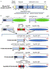
Proteins associated with amyotrophic lateral sclerosis (ALS). (A) Structural and functional organization of TDP-43. NLS—nuclear localization signal;RRM1 and RRM2—RNA recognition motifs 1 and 2, respectively; NES—nuclear export signal; LCD—low complexity domain; QN—QN-rich domain. (B) Structural and functional organization of FUS. Q/G/S/Y-rich—the region rich in glutamine, glycine, serine, and tyrosine; G-rich—the region rich in glycine, RRM—RNA recognition motif, RGG—the motifs containing arginine/glycine/glycine repeats; ZnF—zinc finger domain; NNLS—non-conventional nuclear localization signal. (C) hnRNPA2B1 and yeast chimeric constructs based on this protein: PrLD –prion domain like domain; Core PrLD—core region of PrLD; RRM1 and RRM2—RNA recognition motifs 1 and 2, respectively. Insertion distinguishing hnRNPB1 from hnRNPA2 is shown. Sup35 designations are the same as on Fig. 2 (with the Sup35N domain drawn as a rectangle, as it aggregates when included in a chimeric construct). Numbers indicate amino acid positions in ALS-associated proteins (black font, located under the drawing in a chimeric construct) and Sup35 (red font, located above the drawing in a chimeric construct).
Fig. 10
Fig. 10
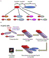
Prion nucleation by mammalian amyloidogenic proteins (MAPs) in yeast. (A) Chimeric prion domains constructed from the Sup35 PrD-containing region fused to Aβ, PrP, αSyn and IAPP, respectively (see text for the description of the regions of amyloidogenic proteins, used in these constructs). (B) Model of de novo prion nucleation by chimeric constructs in the [pin] yeast cells lacking any known pre-existing prions. As shown in Fig. 3, overexpression of Sup35N or Sup35NM alone does not lead to efficient nucleation of the [PSI+] prion in the [pin] cells. Designations of the domains, prion and non-prion forms of Sup35 are the same as on Fig. 2. The region of oligonucleotide repeats (NR) is present, but not shown for the sake of simplicity. See text for detailed description.

References

    1. Aguzzi A, & Lakkaraju AK (2015). Cell biology of prions and prionoids: A status report. Trends in Cell Biology, 26(1), 40–51. 10.1016/j.tcb.2015.08.007. - DOI - PubMed
    1. Aguzzi A, & O’Connor T (2010). Protein aggregation diseases: Pathogenicity and therapeutic perspectives. Nature Reviews. Drug Discovery, 9(3), 237–248. 10.1038/nrd3050. - DOI - PubMed
    1. Ahmed AB, & Kajava AV (2013). Breaking the amyloidogenicity code: Methods to predict amyloids from amino acid sequence. FEBS Letters, 587(8), 1089–1095. 10.1016/j.febslet.2012.12.006. - DOI - PubMed
    1. Ahmed AB, Znassi N, Chateau MT, & Kajava AV (2015). A structure-based approach to predict predisposition to amyloidosis. Alzheimers Dement, 11(6), 681–690. 10.1016/j.jalz.2014.06.007. - DOI - PubMed
    1. Alberti S, Halfmann R, King O, Kapila A, & Lindquist S (2009). A systematic survey identifies prions and illuminates sequence features of prionogenic proteins. Cell, 137(1), 146–158. doi:S0092-8674(09)00266-9 [pii]. 10.1016/j.cell.2009.02.044. - DOI - PMC - PubMed

Publication types

MeSH terms