Review
. 2020 Oct 1:489:144-154.
doi: 10.1016/j.canlet.2020.06.004. Epub 2020 Jun 16.

Hypoxia in bone metastasis and osteolysis

Affiliations
Review

Hypoxia in bone metastasis and osteolysis

Vera M Todd et al. Cancer Lett. .

Abstract

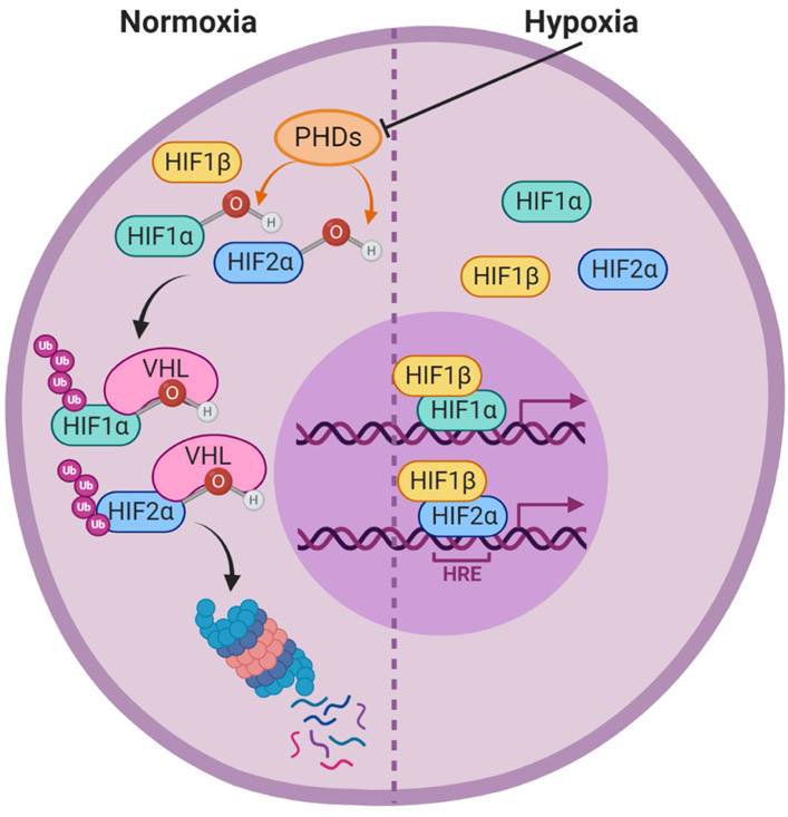
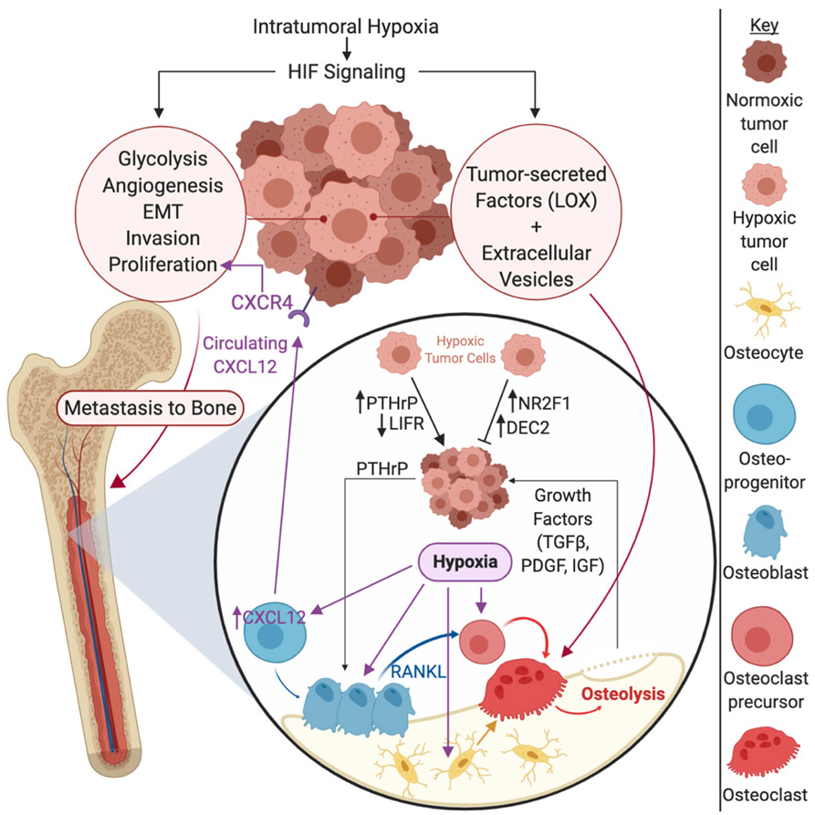
Hypoxia is a common feature in tumors, driving pathways that promote epithelial-to-mesenchymal transition, invasion, and metastasis. Clinically, high levels of hypoxia-inducible factor (HIF) expression and stabilization at the primary site in many cancer types is associated with poor patient outcomes. Experimental evidence suggests that HIF signaling in the primary tumor promotes their dissemination to the bone, as well as the release of factors such as LOX that act distantly on the bone to stimulate osteolysis and form a pre-metastatic niche. Additionally, the bone itself is a generally hypoxic organ, fueling the activation of HIF signaling in bone resident cells, promoting tumor cell homing to the bone as well as osteoclastogenesis. The hypoxic microenvironment of the bone also stimulates the vicious cycle of tumor-induced bone destruction, further fueling tumor cell growth and osteolysis. Furthermore, hypoxia appears to regulate key tumor dormancy factors. Thus, hypoxia acts both on the tumor cells as well as the metastatic site to promote tumor cell metastasis.

Keywords: Cancer; Dormancy; HIF; Oxygen; Vasculature.


Conflict of interest statement

Conflict of interest statement

The authors have nothing to disclose.

Figures

Figure 1.
Figure 1.. Hypoxia-inducible factor signaling pathway.
Under normoxic conditions, prolyl hydroxylase domain containing enzymes (PHDs) hydroxylate the hypoxia inducible factor alpha subunits (HIF1α and HIF2α). These hydroxylations allow the E3 ubituitin ligase von Hippen Lindau (VHL) to ubiquitinate HIF1α and HIF2α, leading to their proteasomal degradation. Under hypoxic conditions, the PHD enzymes are inactive, allowing HIF1α and HIF2α to accumulate in the cytoplasm. The alpha subunits will then dimerize with HIF1β, enter the nucleus, and bind to hypoxia response elements (HREs) in the DNA to act as transcription factors and drive the expression of hypoxia responsive genes.
Figure 2.
Figure 2.. Hypoxia driven factors in the primary tumor and bone metastatic site.
Intratumoral hypoxia in the tumor drives hypoxia inducible factor (HIF) signaling within tumor cells, which drives pro-survival and pro-metastatic processes such as glycolysis, angiogenesis, epithelial-to-mesenchymal transition (EMT), invasion, and tumor cell proliferation. HIF signaling also causes tumor cells to secrete factors such as lysyl oxidase (LOX) and to alter extracellular vesicle production, promoting osteolysis. Once tumor cells reach the bone, a physiologically hypoxic microenvironment, hypoxic tumor cells can be maintained in a dormant state, marked by increased expression of nuclear receptor subfamily 2 group F member 1 (NR2F1) and differentially expressed in chondrocytes 2 (DEC2). On the other hand, hypoxia may drive tumor cells out of dormancy by inducing parathyroid hormone related protein (PTHrP) expression and down regulating leukemia inhibitory factor receptor (LIFR), promoting the establishment of clinically significant metastatic lesions. Hypoxia also promotes the expression of PTHrP, driving the vicious cycle of bone destruction. Hypoxia in the bone also promotes osteolysis by direct stimulation of osteoclastogenic factors by osteoblasts, bone marrow stromal cells, and osteocytes. Intermittent hypoxia also stimulates osteoclastogenesis and osteoclast function. Additionally, hypoxia stimulates CXCL12 expression on osteoblast progenitors and increases circulating CXCL12 levels, which can stimulate the growth and metastasis of primary breast cancer cells through CXCR4, indicating that hypoxia in the bone can exert effects on the primary tumor site as well. Additional abbreviations: TGFβ = transforming growth factor receptor beta; PDGF = platelet derived growth factor; IGF = insulin-like growth factor.

References

    1. Azzi S, Hebda JK, and Gavard J, Vascular permeability and drug delivery in cancers. Front Oncol, 2013. 3: p. 211. - PMC - PubMed
    1. Vaupel P, Hockel M, and Mayer A, Detection and characterization of tumor hypoxia using pO2 histography. Antioxid Redox Signal, 2007. 9(8): p. 1221–35. - PubMed
    1. Semenza GL and Wang GL, A nuclear factor induced by hypoxia via de novo protein synthesis binds to the human erythropoietin gene enhancer at a site required for transcriptional activation. Mol Cell Biol, 1992. 12(12): p. 5447–54. - PMC - PubMed
    1. Wang GL, et al. , Hypoxia-inducible factor 1 is a basic-helix-loop-helix-PAS heterodimer regulated by cellular O2 tension. Proc Natl Acad Sci U S A, 1995. 92(12): p. 5510–4. - PMC - PubMed
    1. Tian H, McKnight SL, and Russell DW, Endothelial PAS domain protein 1 (EPAS1), a transcription factor selectively expressed in endothelial cells. Genes Dev, 1997. 11(1): p. 72–82. - PubMed

Publication types