. 2020 Aug 1;34(15-16):1065-1074.
doi: 10.1101/gad.330050.119. Epub 2020 Jun 19.

Human RTEL1 associates with Poldip3 to facilitate responses to replication stress and R-loop resolution

Affiliations

Human RTEL1 associates with Poldip3 to facilitate responses to replication stress and R-loop resolution

Andrea Björkman et al. Genes Dev. .

Abstract

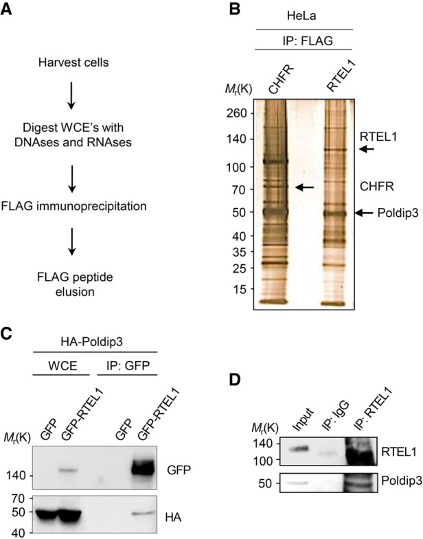
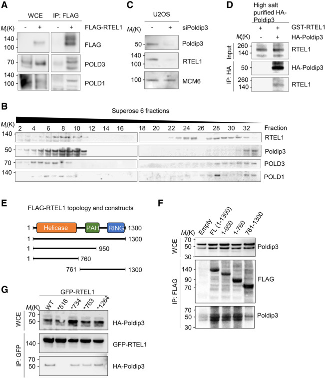
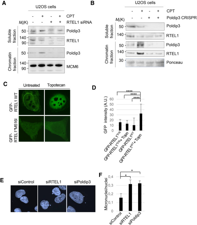
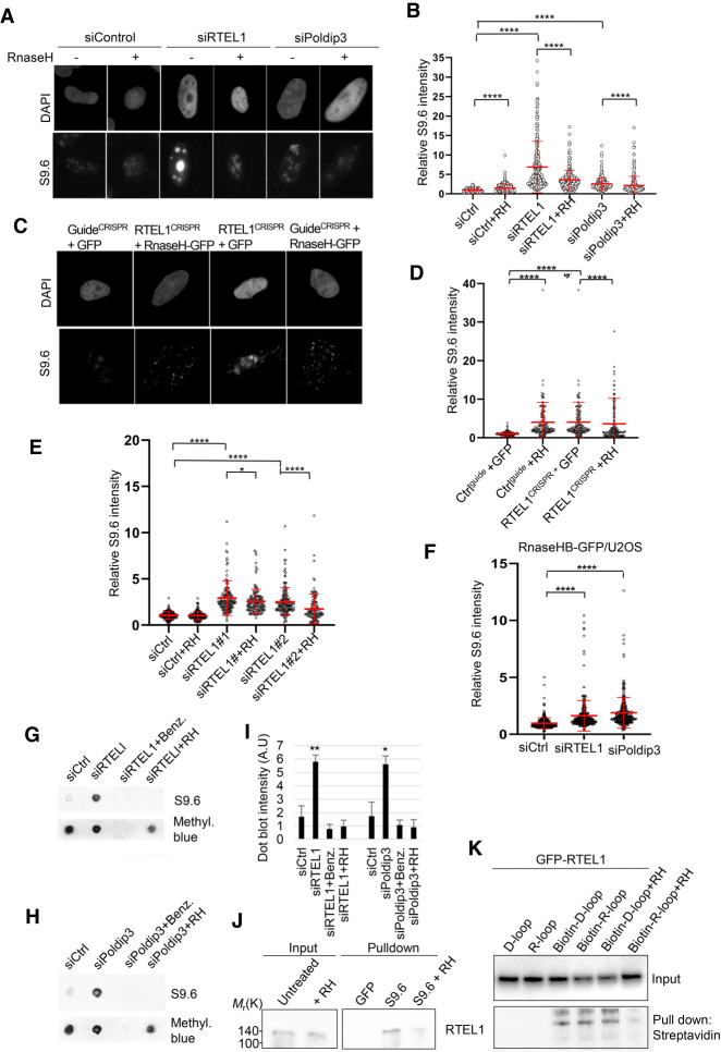
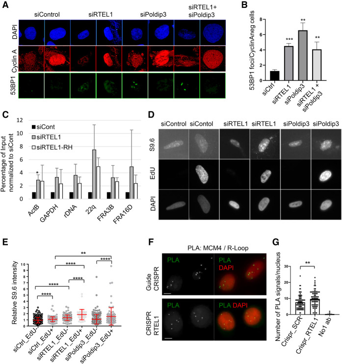
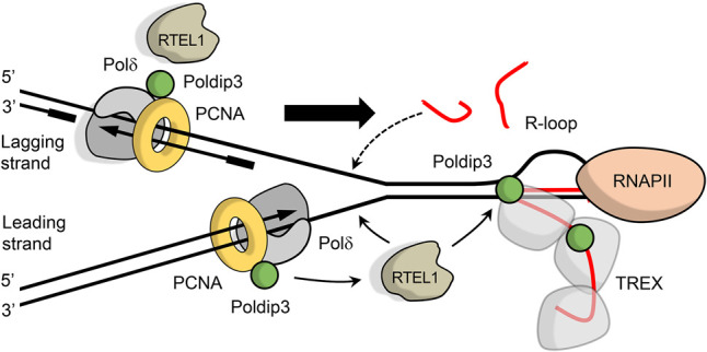
RTEL1 helicase is a component of DNA repair and telomere maintenance machineries. While RTEL1's role in DNA replication is emerging, how RTEL1 preserves genomic stability during replication remains elusive. Here we used a range of proteomic, biochemical, cell, and molecular biology and gene editing approaches to provide further insights into potential role(s) of RTEL1 in DNA replication and genome integrity maintenance. Our results from complementary human cell culture models established that RTEL1 and the Polδ subunit Poldip3 form a complex and are/function mutually dependent in chromatin binding after replication stress. Loss of RTEL1 and Poldip3 leads to marked R-loop accumulation that is confined to sites of active replication, enhances endogenous replication stress, and fuels ensuing genomic instability. The impact of depleting RTEL1 and Poldip3 is epistatic, consistent with our proposed concept of these two proteins operating in a shared pathway involved in DNA replication control under stress conditions. Overall, our data highlight a previously unsuspected role of RTEL1 and Poldip3 in R-loop suppression at genomic regions where transcription and replication intersect, with implications for human diseases including cancer.

Keywords: DNA damage response; DNA repair; DNA:RNA hybrid; Hoyeral-Hreiderson syndrome; POLδ; Poldip3; R-loop; RTEL1; dyskeratosis congenita; helicase; polymerase δ; telomere maintenance.


Figures

Figure 1.
Figure 1.
RTEL1 associates with Poldip3. (A) Schematic flow chart of the Flag affinity purification protocol of Flag-RTEL1. Flag-CHFR or Flag-RTEL1 or expressing HeLa cells were harvested and whole cell lysates were treated with a cocktail of DNases and RNases. Flag-RTEL1 fusion proteins were Flag immunopurified, eluted with Flag peptides, and immunocomplexes resolved by PAGE. (B) Silver staining of resolved proteins from the Flag-RTEL1 immunoprecipitation. Control immunoprecipitation was performed with a Flag-CHFR fusion protein. CHFR was used as control since it, like RTEL1, is a RING finger-containing protein. Flag-CHFR, Flag-RTEL1 and Poldip3 are indicated with arrows. (C) GFP-RTEL1 binds HA-Poldip3. HEK293T cells were cotransfected with HA-Poldip3 and either GFP or GFP-RTEL1 and incubated for 24 h prior to harvest. Extracts were subjected to GFP immunoprecipitation followed by immunoblotting with HA and GFP antibodies. (D) Endogenous RTEL1 binds endogenous Poldip3. U2OS cells were harvested in EBC buffer and cell lysates were treated as in A. Precleared lysates were subjected to immunoprecipitation with either IgG or RTEL1 antibody. RTEL1 immunocomplexes were released from the RTEL1-conjugated resin by treatment with glycine-HCl buffer.
Figure 2.
Figure 2.
RTEL1 binds other Poldδ subunits and directly to Poldip3 through its helicase domain. (A) RTEL1 binds POLD3 and POLD1 subunits of the Polδ complex. Cell extracts from HEK293T cells transfected with Flag-RTEL1 or mock transfected were cleared and subjected to Flag immunoprecipitation with anti-Flag beads followed by PAGE. Proteins were probed by immunoblotting using the indicted antibodies. (B) Gel filtration analysis of HEK293T cell extracts. Proteins were probed by immunoblotting using the indicted antibodies. (C) Codepletion of RTEL1 by Poldip3 silencing. U2OS cells transfected with either control or Poldip3 siRNAs were incubated 4 d and subsequently harvested and proteins immunoblotted using the indicated antibodies. (D) Purified RTEL1 and Poldip3 interact in vitro. Bacterially purified GST-RTEL1 was incubated with high-salt-purified HA (control) or HA-Poldip3 on HA-conjugated beads. Eluded protein complexes were subjected to immunoblot analysis and proteins were probed using either HA or RTEL1 antibodies. (E) Schematic representation of Flag-RTEL1 deletion constructs. (F) RTEL1 binds Poldip3 primarily via its 4FeS helicase domain. HEK293T cells were transfected with Flag-RTEL1 full-length or deletion constructs spanning the indicated regions of RTEL1 to determine the Poldip3-binding site. The cell lysates were subjected to Flag immunoprecipitation followed by 3xFlag peptide elution and, subsequently, proteins were subjected to immunoblotting with Flag or Poldip3 antibodies. (G) GFP pull-down analysis of mutated GFP-RTEL1. HEK293T cells were cotransfected with HA-Poldip3 and either GFP-RTEL1 WT or GFP-RTEL1 containing the Hoyeraal-Hreidarsson syndrome mutations at M516I, L734R, G763V, or R1264H. Cell extracts were subjected to GFP immunoprecipitation followed by immunoblotting with GFP or HA antibodies.
Figure 3.
Figure 3.
RTEL1 and Poldip3 bind chromatin in a mutually dependent manner after CPT treatment. (A) RTEL1 is required for chromatin recruitment of Poldip3 after CPT treatment. Whole cell extracts of WT or RTEL1 silenced U2OS cells were treated with CPT for 24 h, harvested, and split into soluble and chromatin fractions and chromatin bound proteins were released by acid extraction. Proteins were detected by immunoblotting with the indicated antibodies. (B) Poldip3 is required for chromatin recruitment of RTEL1 after CPT treatment. Whole-cell extracts of WT or Poldip3 CRISPR silenced U2OS cells were treated with CPT for 24 h, harvested, and split into soluble and chromatin fractions and chromatin-bound proteins were released by acid extraction. Proteins were detected by immunoblotting with the indicated antibodies. (C) GFP-RTEL1 WT or M516I mutated GFP-RTEL1 nuclear localization. U2OS cells stably expressing GFP-RTEL1 WT or M516 mutated GFP-RTEL1 were left untreated or treated with topotecan for 24 h. (D) Quantification of nuclear localization of GFP-RTEL1in U2OS cells stably expressing GFP-RTEL1 WT or GFP-RTEL1*M516I. Nuclear minus cytoplasmic staining is shown. (****) P < 0.0001, two-tailed Mann-Whitney test. (E) Increased micronuclei formation in cells depleted of RTEL1 or Poldip3. Representative images showing DAPI staining in U2OS cells treated with control, RTEL1, or Poldip3 siRNA. (F) Quantification of micronuclei in U2OS cells treated with control, RTEL1, or Poldip3 siRNAs. Mean and SD of three independent experiments are shown. (*) P < 0.05, Student's t-test.
Figure 4.
Figure 4.
Increased accumulation of R loops in RTEL1- or Poldip3-depleted cells. (A) Increased accumulation of R loops in cells depleted of RTEL1 or Poldip3. Representative images of DAPI and S9.6 fluorescence staining in U2OS cells treated with control, RTEL1, or Poldip3 siRNAs. (B) Quantification of nuclear S9.6 fluorescence intensity in U2OS cells treated with control, RTEL1, or Poldip3 siRNA. Cells conditionally overexpressing GFP-RNase H (RH) with doxycline were used. (C) Increased amount of R-loops in RTEL1 knockout U2OS cells. U2OS cells transiently transfected with CRISPR–CAS9 guides against RTEL1 or control guide and RNase H or GFP. (D) Quantification of nuclear S9.6 fluorescence intensity in U2OS cells transiently transfected with CRISPR-CAS9 guides against RTEL1 or control guide and RNase H or GFP. (E) Increased accumulation of R-loops in U2OS cells depleted of RTEL1 using two different RTEL1 siRNAs. Quantification of nuclear S9.6 fluorescence intensity in U2OS cells conditionally overexpressing GFP-RNase H (RH) treated with control or RTEL1 siRNAs. (F) Increased GFP-RNase H/R-loop binding domain recruitment to nucleus in cells depleted of RTEL1 or Poldip3. Quantification of GFP fluorescence intensity in U2OS-RNase HB-GFP cells treated with control, RTEL1, or Poldip3 siRNA. (G) R-loop dot blot analysis. U2OS cells were treated with either control, RTEL1, or Poldip3 siRNA followed by a 3-d incubation. Genomic DNA was isolated from cell lysates, bulk RNA digested with RNase A, and DNA was subsequently hand spotted on activated nylon membranes as indicated. DNA digested with the nuclease benzonase (Benz.) or RNase H (RH) was used as a control. (H) R-loop dot blot analysis. U2OS cells were treated with either control or Poldip3 siRNA followed by a 3-d incubation. Genomic DNA was isolated from cell lysates, bulk RNA digested with RNase A, and DNA was subsequently hand-spotted on activated nylon membranes as indicated. DNA digested with the nuclease benzonase (Benz.) or RNase H (RH) was used as a control. (I) Quantification of G and H. Mean and SD are shown. (*) P < 0.05; (**) P < 0.01, Student's t-test. (J) RTEL1 binds to R-loops. U2OS cells left untreated or treated with CPT for 24 h were harvested and R-loops were immunoprecipitated from cleared nuclear extracts with the S9.6 antibody. Nuclear extracts from cells treated with RNase H (RH) was used as a control. (K) RTEL1 interacts with R-loops and D-loops in vitro. Extracts of U2OS cells stably expressing GFP-RTEL1 WT were incubated with biotin-coupled R-loop or D-loop hybrids immobilized on streptavidin beads, and GFP-RTEL1 was immunoblotted with GFP antibody. Cell extracts incubated with purified RNase H (RH) was used as a control. Mean and SD are plotted. (B,D,E,F) (*) P < 0.05; (****) P < 0.0001, two-tailed Mann-Whitney test. Data from three or more independent experiments are combined and immunofluorescence intensities are normalized to siCtrl.
Figure 5.
Figure 5.
R loops accumulate at specific genomic loci and associate with replication after RTEL1 depletion. (A) Increased 53BP1 bodies in Cyclin A-negative U2OS cells depleted of RTEL1, Poldip3, or RTEL1 and Poldip3. Representative images showing DAPI and 53BP1 staining in U2OS cells treated with control, RTEL1, Poldip3, or RTEL1 and Poldip3 siRNA. (B) Quantification of A. The average of three independent experiments are shown. (**) P < 0.01; (***) P < 0.001, Student's t-test. (C) DNA:RNA hybrids accumulate at commonly expressed genes (GAPDH and ActB), common fragile sites (FRA3B and FRA16D), telomeres (22q) and rDNA in U2OS cells depleted of RTEL1. Results are obtained with qPCR from DNA samples captured by DNA–RNA immunoprecipitation (DRIP) from siCont or siRTEL1 U2OS samples untreated or treated with RNase H (RH). Percentage of input normalized to siCont from three experiments are shown. (*) P < 0.01, Student's t-test. (D) Increased amount of R loops in EdU-positive RTEL1- or Poldip3-depleted cells. Representative images of S9.6, EdU, and DAPI staining in RTEL1- and Poldip3-depleted cells. (E) Quantification of S9.6 nuclear fluorescence intensity in control, RTEL1 or Poldip3 siRNA-treated EdU-positive or EdU-negative U2OS cells. Values normalized to siCtrl. Mean and SD are plotted (**) P < 0.01; (****) P < 0.0001, two-tailed Mann-Whitney test. (F) Proximity ligation assay (PLA) of S9.6 and MCM4 after RTEL1 knockout. HeLa cells sorted (GFP) 48 h after transfection with either CRISPR–Cas9 against RTEL1 or CRISPR control and put on slides. PLA revealing MCM4 and R-loops. (G) Quantification of F. (SCR) Scrambled control guide. (**) P < 0.01, Mann-Whitney test.
Figure 6.
Figure 6.
Cartoon showing our model of the role of RTEL1 and Poldip3 in R-loop resolution. RTEL1 associates with the replisome, here represented by Polδ (gray) and PCNA (yellow) via Poldip3 and/or PCNA, as it replicates DNA. As the replication fork encounters the transcription machinery, RTEL1 might resolve R-loops that block the replication fork and/or the R-loops provoked by stalling of replication (shown by arrows from RTEL1). (RNAPII) RNA polymerase II. The black lines represent single-stranded DNA and the red lines depict RNA. The big arrow indicates the direction of the replication fork.

References

    1. Ballew BJ, Joseph V, De S, Sarek G, Vannier JB, Stracker T, Schrader KA, Small TN, O'Reilly R, Manschreck C, et al. 2013a. A recessive founder mutation in regulator of telomere elongation helicase 1, RTEL1, underlies severe immunodeficiency and features of Hoyeraal Hreidarsson syndrome. PLoS Genet 9: e1003695 10.1371/journal.pgen.1003695 - DOI - PMC - PubMed
    1. Ballew BJ, Yeager M, Jacobs K, Giri N, Boland J, Burdett L, Alter BP, Savage SA. 2013b. Germline mutations of regulator of telomere elongation helicase 1, RTEL1, in dyskeratosis congenita. Hum Genet 132: 473–480. 10.1007/s00439-013-1265-8 - DOI - PMC - PubMed
    1. Barber LJ, Youds JL, Ward JD, McIlwraith MJ, O'Neil NJ, Petalcorin MI, Martin JS, Collis SJ, Cantor SB, Auclair M, et al. 2008. RTEL1 maintains genomic stability by suppressing homologous recombination. Cell 135: 261–271. 10.1016/j.cell.2008.08.016 - DOI - PMC - PubMed
    1. Boubakri H, de Septenville AL, Viguera E, Michel B. 2010. The helicases DinG, Rep and UvrD cooperate to promote replication across transcription units in vivo. EMBO J 29: 145–157. 10.1038/emboj.2009.308 - DOI - PMC - PubMed
    1. Deng Z, Glousker G, Molczan A, Fox AJ, Lamm N, Dheekollu J, Weizman OE, Schertzer M, Wang Z, Vladimirova O, et al. 2013. Inherited mutations in the helicase RTEL1 cause telomere dysfunction and Hoyeraal-Hreidarsson syndrome. Proc Natl Acad Sci 110: E3408-16. - PMC - PubMed

Publication types

LinkOut - more resources