. 2020 Aug 18;86(17):e00971-20.
doi: 10.1128/AEM.00971-20. Print 2020 Aug 18.

Insights into the Maturation of Pernisine, a Subtilisin-Like Protease from the Hyperthermophilic Archaeon Aeropyrum pernix

Affiliations

Insights into the Maturation of Pernisine, a Subtilisin-Like Protease from the Hyperthermophilic Archaeon Aeropyrum pernix

Miha Bahun et al. Appl Environ Microbiol. .

Abstract

Pernisine is a subtilisin-like protease that was originally identified in the hyperthermophilic archaeon Aeropyrum pernix, which lives in extreme marine environments. Pernisine shows exceptional stability and activity due to the high-temperature conditions experienced by A. pernix Pernisine is of interest for industrial purposes, as it is one of the few proteases that has demonstrated prion-degrading activity. Like other extracellular subtilisins, pernisine is synthesized in its inactive pro-form (pro-pernisine), which needs to undergo maturation to become proteolytically active. The maturation processes of mesophilic subtilisins have been investigated in detail; however, less is known about the maturation of their thermophilic homologs, such as pernisine. Here, we show that the structure of pro-pernisine is disordered in the absence of Ca2+ ions. In contrast to the mesophilic subtilisins, pro-pernisine requires Ca2+ ions to adopt the conformation suitable for its subsequent maturation. In addition to several Ca2+-binding sites that have been conserved from the thermostable Tk-subtilisin, pernisine has an additional insertion sequence with a Ca2+-binding motif. We demonstrate the importance of this insertion for efficient folding and stabilization of pernisine during its maturation. Moreover, analysis of the pernisine propeptide explains the high-temperature requirement for pro-pernisine maturation. Of note, the propeptide inhibits the pernisine catalytic domain more potently at high temperatures. After dissociation, the propeptide is destabilized at high temperatures only, which leads to its degradation and finally to pernisine activation. Our data provide new insights into and understanding of the thermostable subtilisin autoactivation mechanism.IMPORTANCE Enzymes from thermophilic organisms are of particular importance for use in industrial applications, due to their exceptional stability and activity. Pernisine, from the hyperthermophilic archaeon Aeropyrum pernix, is a proteolytic enzyme that can degrade infective prion proteins and thus has a potential use for disinfection of prion-contaminated surfaces. Like other subtilisin-like proteases, pernisine needs to mature through an autocatalytic process to become an active protease. In the present study, we address the maturation of pernisine and show that the process is regulated specifically at high temperatures by the propeptide. Furthermore, we demonstrate the importance of a unique Ca2+-binding insertion for stabilization of mature pernisine. Our results provide a novel understanding of thermostable subtilisin autoactivation, which might advance the development of these enzymes for commercial use.

Keywords: Aeropyrum pernix; Ca2+ binding; maturation; pernisine; propeptide; subtilisin.


Figures

FIG 1
FIG 1
Schematic representation of pro-pernisine primary structure and predicted Ca2+ binding sites. (Top) The numbers above the diagram define the amino acid numbering. The signal sequence (SIG.) starts with Met1, the propeptide starts with Ala27, and the catalytic domain starts with Ala95. The unique insertion in pernisine (violet bar) is compared to the corresponding regions of Tk-subtilisin and subtilisin E. The consensus Ca2+-binding motif in the insertion is underlined, and the predicted Ca2+-binding residues are highlighted in violet. (Bottom) Defined amino acid sequences of the insertions (IS1 and IS2) and the predicted Ca2+-binding sites (Ca1 to Ca7) inferred by the alignment of pernisine (UniProt accession no. Q9YFI3), Tk-subtilisin (UniProt accession no. P58502), and subtilisin E (UniProt accession no. P04189). The amino acids shown to interact with Ca2+ in Tk-subtilisin (18) and the corresponding residues in pernisine and subtilisin E are highlighted in violet. Black shading, identical amino acids; gray shading, similar amino acids. Clustal Omega (41) and BoxShade tools were used for the alignments.
FIG 2
FIG 2
Pro-pernisine variants for maturation assays. (A) SDS-PAGE analysis of pro-Per, pro-PerΔCa, and pro-PerΔIS isolated by affinity chromatography. The positions of the unprocessed pro-pernisine, pro-pernisineΔCa, and pro-pernisineΔIS; the autoprocessed forms of pro-pernisine and pro-pernisineΔIS (pro-Per* and pro-PerΔIS*, respectively); and the propeptide are indicated with arrows. Note that the autoprocessed form of pro-pernisineΔCa was not present. Degraded forms of these proteins are also marked. The N termini of the SDS-PAGE bands corresponding to pro-pernisine and pro-pernisine* were determined to be Ala27 and Ala95, respectively. Lanes M, protein weight markers, with molecular masses shown next to the gels. (B) Schematic representation of the unprocessed pernisine variants (pro-Per, pro-PerΔCa, and pro-PerΔIS) and their autoprocessed forms (pro-Per* and pro-PerΔIS*). The unique insertion is represented by the violet bars. For pro-pernisineΔCa, the mutations in the predicted Ca2+-binding motif within the insertion (D134A/N136A/D138A) are indicated in red. Amino acid numbering is as in Fig. 1. For the autoprocessed forms, the cleaved scissile bond between the propeptide and the catalytic domain is indicated by a dashed line. Note that the signal sequence of the OmpA protein and the native pernisine signal sequence were not present after isolation from the periplasm, as confirmed by N-terminal sequencing of pro-Per. The 10× histidine tags (His10) are also indicated.
FIG 3
FIG 3
Maturation of pro-pernisine and its variants with the unique Ca2+ site mutated (pro-pernisineΔCa) and the insertion deleted (pro-pernisineΔIS) at different CaCl2 concentrations. Pro-pernisine, pro-pernisineΔCa, and pro-pernisineΔIS (40 μg/ml) were incubated in the absence of CaCl2 and with 100 μM, 1 mM, and 10 mM CaCl2 at 90°C for the indicated times. Their maturation was analyzed using SDS-PAGE and azocasein assays, as described in Materials and Methods. Lanes M, protein weight markers, with molecular masses shown next to the gels. The arrows indicate the unprocessed form (gray), the autoprocessed/mature form (black), and the propeptide (dashed). The proteolytic activities were measured at the indicated times at 90°C in triplicate and are plotted below the gels, in alignment with their corresponding lanes. The error bars indicate standard deviations. The CaCl2 concentration in the azocasein assays was 1 mM.
FIG 4
FIG 4
Maturation of pro-pernisine (A) and its variants with the unique Ca2+ site mutated (B) and with the insertion deleted (C) at different CaCl2-to-protein molar ratios. The proteins were incubated at 90°C for 6 h at the indicated molar ratios of CaCl2 to protein and analyzed using SDS-PAGE and azocasein assays, as described in Materials and Methods. The molar concentration of the target protein (1 μM) was estimated from the mass concentration (40 μg/ml). Lanes M, molecular masses of the protein markers, as shown next to the gels. The arrows indicate the unprocessed form (gray), the autoprocessed/mature form (black), and the propeptide (dashed). The proteolytic activities at the indicated ratios were recorded at 90°C in triplicate and are plotted below the gels, in alignment with their corresponding lanes. The error bars indicate standard deviations. The CaCl2 concentration in azocasein assays was 1 mM.
FIG 5
FIG 5
Maturation of pro-pernisine and its variants with the unique Ca2+ site mutated (pro-pernisineΔCa) and with the insertion deleted (pro-pernisineΔIS) at different temperatures, as indicated. Pro-pernisine, pro-pernisineΔCa, and pro-pernisineΔIS (40 μg/ml) were incubated at 60°C, 70°C, and 80°C for the indicated times in the presence of 10 mM CaCl2. Their maturation was analyzed using SDS-PAGE and azocasein assays, as described in Materials and Methods. Lanes M, molecular masses of the protein markers, as shown next to the gels. The arrows indicate the unprocessed form (gray), the autoprocessed/mature form (black), and the propeptide (dashed). The proteolytic activities at the indicated times were measured at 90°C in triplicate, with the data plotted below the gels, in alignment with their corresponding lanes. The error bars indicate standard deviations. The CaCl2 concentration in the azocasein assays was 1 mM.
FIG 6
FIG 6
Active-site mutants of the pro-pernisine variants. (A) Analysis of pro-pernisineS355A (pro-PerS355A) purity after IMAC and after the final purification step by SDS-PAGE and Western blotting. Lanes M, molecular masses of the protein markers, as shown next to the gels. The arrows (right) indicate the bands that correspond to the unprocessed pro-pernisineS355A and the two N-terminally truncated variants of pro-pernisineS355A (Asn136–Ser430 and Ala146–Ser430). These bands were subjected to N-terminal sequencing after IMAC. The band corresponding to the target pro-pernisineS355A was also subjected to N-terminal sequencing after the final purification step. (B) Schematic representations of the isolated unprocessed pro-pernisine variants that were used for folding analyses: pro-pernisineS355A, the unprocessed pro-pernisineS355A; pro-pernisineΔCaS355A (pro-PerΔCaS355A), the unprocessed pro-pernisineΔCaS355A; and pro-pernisineΔISS355A (pro-PerΔISS355A), the unprocessed pro-pernisineΔISS355A. The unique insertion and the mutations in the Ca2+-binding motif are indicated as in Fig. 2. The Ser355→Ala355 substitution in the active site is indicated by the asterisk (S355A). Amino acid numbering is as in Fig. 1. Note that the signal sequence of the OmpA protein and the native pernisine sequence were not present after isolation from the periplasm.
FIG 7
FIG 7
Folding analyses of the pro-pernisine active-site mutant (pro-pernisineS355A) and its variants with the unique Ca2+-binding site mutated (pro-pernisineΔCaS355A) or insertion deleted (pro-pernisineΔISS355A). (A to C) Far-UV CD (A), near-UV CD (B), and tryptophan fluorescence (C) spectra. Red lines, pro-pernisineS355A; black lines, pro-pernisineΔCaS355A; gray lines, pro-pernisineΔISS355A; dashed lines, without CaCl2; solid lines, with CaCl2 (CaCl2-to-protein molar ratio, 200:1). (C) The fluorescence spectra of pro-pernisineS355A, pro-pernisineΔCaS355A, and pro-pernisineΔISS355A were normalized to their respective spectra measured in the absence of CaCl2. (D to F) The molar ellipticities at 200 nm (D) and 280 nm (E) and the ratios of the fluorescence intensities at 330 nm and 350 nm (F) were plotted as a function of the CaCl2-to-protein ratio. Red, pro-pernisineS355A; black, pro-pernisineΔCaS355A; gray, pro-pernisineΔISS355A. (G to I) SDS-PAGE analysis of the Ca2+-induced folding of pro-pernisineS355A (G), pro-pernisineΔCaS355A (H), and pro-pernisineΔIS S355A (I). The ratios of CaCl2 to protein are as indicated above the lanes. Gray arrows, Ca2+-free form of pro-pernisineS355A, pro-pernisineΔCaS355A, or pro-pernisineΔISS355A; black arrows, Ca2+-bound form of pro-pernisineS355A, pro-pernisineΔCaS355A, or pro-pernisineΔISS355A; dashed arrows, Ca2+-free form of N-terminally truncated pro-pernisineS355A or pro-pernisineΔCaS355A. Lanes where the sample was precipitated with trichloroacetic acid prior to SDS-PAGE are indicated with black dots.
FIG 8
FIG 8
Conformational stability of the pro-pernisine active-site mutant (pro-pernisineS355A) and its variants with the unique Ca2+-binding site mutated (pro-pernisineΔCaS355A) or with the insertion deleted (pro-pernisineΔISS355A). The far-UV CD (A) and near-UV CD (B) spectra were measured at a CaCl2-to-protein molar ratio of 200:1 at 20°C (solid lines) and at 90°C (dashed lines). Red lines, pro-pernisineS355A; black lines, pro-pernisineΔCaS355A; gray lines, pro-pernisineΔISS355A.
FIG 9
FIG 9
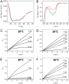
Characterization of propeptide conformation and inhibitory activity. (A and B) The far-UV CD (A) and near-UV CD (B) spectra of the propeptide were recorded at 20°C (black lines) and 90°C (red lines). (C to F) The activities of the mature pernisine (2 nM) at the indicated concentrations of the propeptide were recorded at 20°C (C and D) and 90°C (E and F) using Suc-AAPF-pNA (0.3 mM) as a substrate, as described in Materials and Methods. The propeptide was either mixed with Suc-AAPF-pNA prior to addition of the mature pernisine (C and E) or incubated with the mature pernisine prior to addition of Suc-AAPF-pNA (D and F).
FIG 10
FIG 10
Schematic model for pro-pernisine maturation. The propeptide (violet) is connected to the catalytic domain (blue) via the peptide linker (black line), which contains the scissile bond. The linker is composed of the C-terminal region of the propeptide and the N-terminal region of the catalytic domain. The active site on the catalytic domain is represented by an orange asterisk. Disordered/destabilized domains are bordered by dotted lines, whereas folded domains are bordered by solid lines. (A) In the absence of Ca2+, the disordered pro-pernisine undergoes fast autoprocessing (i.e., cleavage of the scissile bond) into the noncovalent complex of the propeptide-catalytic domain that dissociates reversibly. At 20°C, the dissociated propeptide is resistant to degradation by the catalytic domain. At 90°C, the autoprocessed complex is subjected to autolysis. (B) In the presence of Ca2+, the unprocessed pro-pernisine adopts a folded conformation. Autoprocessing of this Ca2+-bound pro-pernisine is slow. At 20°C, the noncovalent complex of the propeptide-catalytic domain dissociates more readily than at 90°C, as shown by the arrows. The dissociated propeptide is destabilized and degraded by the Ca2+-bound catalytic domain only at 90°C, which leads to formation of the mature pernisine.

References

    1. Siezen RJ, de Vos WM, Leunissen JA, Dijkstra BW. 1991. Homology modelling and protein engineering strategy of subtilases, the family of subtilisin-like serine proteinases. Protein Eng 4:719–737. doi:10.1093/protein/4.7.719. - DOI - PubMed
    1. Siezen RJ, Leunissen J. 1997. Subtilases: the superfamily of subtilisin-like serine proteases. Protein Sci 6:501–523. doi:10.1002/pro.5560060301. - DOI - PMC - PubMed
    1. Yabuta Y, Takagi H, Inouye M, Shinde U. 2001. Folding pathway mediated by an intramolecular chaperone: propeptide release modulates activation precision of pro-subtilisin. J Biol Chem 276:44427–44434. doi:10.1074/jbc.M107573200. - DOI - PubMed
    1. Pantoliano MW, Whitlow M, Wood JF, Rollence ML, Finzel BC, Gilliland GL, Poulos TL, Bryan PN. 1988. The engineering of binding affinity at metal ion binding sites for the stabilization of proteins: subtilisin as a test case. Biochemistry 27:8311–8317. doi:10.1021/bi00422a004. - DOI - PubMed
    1. Pantoliano MW, Whitlow M, Wood JF, Dodd SW, Hardman KD, Rollence ML, Bryan PN. 1989. Large increases in general stability for subtilisin BPN′ through incremental changes in the free energy of unfolding. Biochemistry 28:7205–7213. doi:10.1021/bi00444a012. - DOI - PubMed

Publication types

LinkOut - more resources