. 2020 Aug 14;295(33):11572-11583.
doi: 10.1074/jbc.RA120.012596. Epub 2020 Jun 19.

An astrocyte cell line that differentially propagates murine prions

Affiliations

An astrocyte cell line that differentially propagates murine prions

Waqas Tahir et al. J Biol Chem. .

Abstract

Prion diseases are fatal infectious neurodegenerative disorders in human and animals caused by misfolding of the cellular prion protein (PrPC) into the pathological isoform PrPSc Elucidating the molecular and cellular mechanisms underlying prion propagation may help to develop disease interventions. Cell culture systems for prion propagation have greatly advanced molecular insights into prion biology, but translation of in vitro to in vivo findings is often disappointing. A wider range of cell culture systems might help overcome these shortcomings. Here, we describe an immortalized mouse neuronal astrocyte cell line (C8D1A) that can be infected with murine prions. Both PrPC protein and mRNA levels in astrocytes were comparable with those in neuronal and non-neuronal cell lines permitting persistent prion infection. We challenged astrocytes with three mouse-adapted prion strains (22L, RML, and ME7) and cultured them for six passages. Immunoblotting results revealed that the astrocytes propagated 22L prions well over all six passages, whereas ME7 prions did not replicate, and RML prions replicated only very weakly after five passages. Immunofluorescence analysis indicated similar results for PrPSc Interestingly, when we used prion conversion activity as a readout in real-time quaking-induced conversion assays with RML-infected cell lysates, we observed a strong signal over all six passages, comparable with that for 22L-infected cells. These data indicate that the C8D1A cell line is permissive to prion infection. Moreover, the propagated prions differed in conversion and proteinase K-resistance levels in these astrocytes. We propose that the C8D1A cell line could be used to decipher prion strain biology.

Keywords: C8D1A; Creutzfeldt–Jakob disease; astrocyte; astrocytes; bovine spongiform encephalopathy; neurodegeneration; neurodegenerative disease; prion; prion disease; prion infection; prion strain; protein misfolding; scrapie.


Conflict of interest statement

Conflict of interest—Conflict of interest statement: The authors declare that they have no conflicts of interest with the contents of this article.

Figures
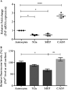
Figure 1.
Figure 1.
Expression of PrPC in C8D astrocytes as compared with other neuronal and non-neuronal cell lines. A, mRNA expression of Prnp gene in four different cell lines. Relative fold change for PrP mRNA expression was measured by RT-PCR. 1 × 10⁶ cells were plated in triplicate, and after 4 days of culture, total RNA was extracted and reverse-transcribed. cDNA was used for quantitative real-time PCR using SYBR. Relative mRNA expression was normalized with actin and calculated using the ΔΔCt method. *, p value ≤ 0.05 and ***, p value ≤ 0.0001 when compared with astrocytes using ANOVA (Dunnett's multiple comparisons) in GraphPad Prism. Error bars represent averages ± S.E. B, PrPC surface levels in four different cell lines. Surface PrPC was analyzed by FACS after staining with mAb 4H11 and Alexa 405–conjugated secondary antibody. Relative mean fluorescence values of N2a, MEF, and CAD5 were compared with the average fluorescence value of astrocytes, which was set to 100%. Statistical analysis was performed by one-way ANOVA and Dunnett's multiple comparison tests using GraphPad Prism. *, p value ≤ 0.05; **, p value ≤ 0.01. Error bars represent averages ± S.E.
Figure 2.
Figure 2.
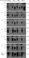
Levels of PrPSc in C8D astrocytes infected with 22L, RML, and ME7 prions. Immunoblots showing PrPSc levels in astrocytes infected with 22L, RML, and ME7 prions as compared with mock-infected astrocytes (before and after PK digestion). The cells from passage 0–6 were lysed and subjected to PK digestion (+PK) or no PK digestion (−PK). The lysates were immunoblotted and probed with mAb 4H11. α-Tubulin was used as loading control.
Figure 3.
Figure 3.
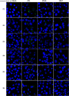
Immunofluorescence analysis of PrPSc aggregates in C8D astrocytes infected with 22L, RML, and ME7 prions. Representative images of PrPSc immunofluorescence staining in astrocytes infected with 22L, RML, and ME7 prion strains compared with mock-infected astrocytes from passages 1–6. Astrocytes were treated with 6 m guanidine hydrochloride prior to antibody incubation to denature PrPC and retrieve PrPSc epitopes. PrPSc was stained with 4H11 as anti PrP antibody (green), and the nuclei were counterstained with 4′,6′-diamino-2-phenylindole (blue). The cells were visualized by confocal laser scanning microscopy. Scale bars represent 20 μm.
Figure 4.
Figure 4.
Prion conversion activity in C8D astrocytes infected with 22L, RML, and ME7 prions. RT-QuIC assay was performed to determine prion conversion activity in astrocytes infected with 22L, RML, and ME7 prion strains as compared with mock-infected astrocytes. Mouse recombinant PrP was used as substrate. Each reaction was set up in quadruplicate with 2 μl of cell lysate (dilution 10-1 is shown). The average increase of thioflavin-T fluorescence of quadruplicates is plotted as a function of time. The y axis represents relative fluorescent units (RFU), and the x axis shows the time in hours. A–F, RT-QuIC analysis of prion-infected astrocytes from passages 1–6 is shown.
Figure 5.
Figure 5.
Stable single-cell clones infected with 22L prions. A, immunoblot analysis showing PrP before (lanes 1 and 2) and after PK digestion (lanes 3 and 4) in noninfected astrocytes and astrocyte clone N31 persistently infected with 22L prions. Tubulin was used as loading control. B, RT-QuIC analysis of the persistently infected astrocyte clone N31. The y axis represents relative fluorescent units (RFU), and the x axis shows the time in hours. Noninfected astrocyte cell lysate was used as negative control, and ScN2a cell lysate was used as positive control. The cells were frozen after passaging for over 6 months for 20 passages after 22L infection. The cells were thawed and cultured again. Immunoblotting and RT-QuIC data represents passage 2 after thawing.

Similar articles

Cited by

References

    1. Billette de Villemeur T., Deslys J. P., Pradel A., Soubrié C., Alpérovitch A., Tardieu M., Chaussain J. L., Hauw J. J., Dormont D., Ruberg M., and Agid Y. (1996) Creutzfeldt–Jakob disease from contaminated growth hormone extracts in France. Neurology 47, 690–695 10.1212/wnl.47.3.690 - DOI - PubMed
    1. Will R. G., Ironside J. W., Zeidler M., Cousens S. N., Estibeiro K., Alperovitch A., Poser S., Pocchiari M., Hofman A., and Smith P. G. (1996) A new variant of Creutzfeldt–Jakob disease in the UK. Lancet 347, 921–925 10.1016/S0140-6736(96)91412-9 - DOI - PubMed
    1. Raymond G. J., Hope J., Kocisko D. A., Priola S. A., Raymond L. D., Bossers A., Ironside J., Will R. G., Chen S. G., Petersen R. B., Gambetti P., Rubenstein R., Smits M. A., Lansbury P. T., and Caughey B. (1997) Molecular assessment of the potential transmissibilities of BSE and scrapie to humans. Nature 388, 285–288 10.1038/40876 - DOI - PubMed
    1. Hsiao K., Baker H. F., Crow T. J., Poulter M., Owen F., Terwilliger J. D., Westaway D., Ott J., and Prusiner S. B. (1989) Linkage of a prion protein missense variant to Gerstmann–Sträussler syndrome. Nature 338, 342–345 10.1038/338342a0 - DOI - PubMed
    1. Prusiner S. B. (1989) Scrapie prions. Annu. Rev. Microbiol. 43, 345–374 10.1146/annurev.mi.43.100189.002021 - DOI - PubMed

Publication types