. 2020 Jun 19;7(1):184.
doi: 10.1038/s41597-020-0508-5.

A clinically and genomically annotated nerve sheath tumor biospecimen repository

Affiliations

A clinically and genomically annotated nerve sheath tumor biospecimen repository

Kai Pollard et al. Sci Data. .

Abstract

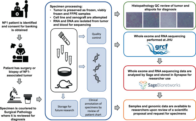
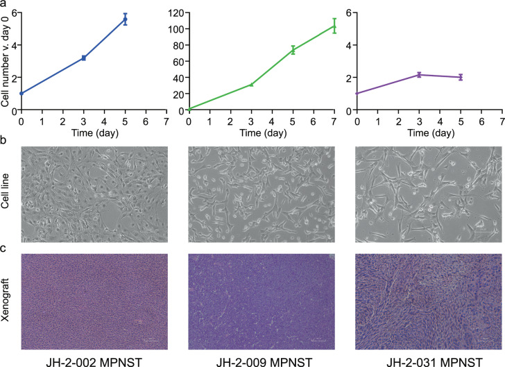
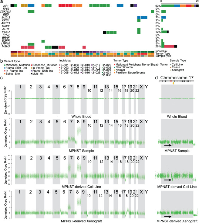
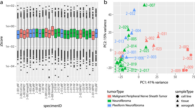
Nerve sheath tumors occur as a heterogeneous group of neoplasms in patients with neurofibromatosis type 1 (NF1). The malignant form represents the most common cause of death in people with NF1, and even when benign, these tumors can result in significant disfigurement, neurologic dysfunction, and a range of profound symptoms. Lack of human tissue across the peripheral nerve tumors common in NF1 has been a major limitation in the development of new therapies. To address this unmet need, we have created an annotated collection of patient tumor samples, patient-derived cell lines, and patient-derived xenografts, and carried out high-throughput genomic and transcriptomic characterization to serve as a resource for further biologic and preclinical therapeutic studies. In this work, we release genomic and transcriptomic datasets comprised of 55 tumor samples derived from 23 individuals, complete with clinical annotation. All data are publicly available through the NF Data Portal and at http://synapse.org/jhubiobank.


Conflict of interest statement

The authors declare no competing interests.

Figures

Fig. 1
Fig. 1
Overview of the Johns Hopkins University NF1 biospecimen repository.
Fig. 2
Fig. 2
Characterization of patient-derived models. (a,b) Cells were cultured under standard conditions until the emergence of a consistently replicating population. (a) Measurement of cell growth rate and calculation of doubling time, shown as a percentage increase over day 0. (b) 10x photomicrograph of cultured cells at logarithmic growth phase. (c) Tumor fragments from freshly acquired specimens were implanted subcutaneously into mice; mice were monitored until the development of a palpable tumor. H&E images from three representative MPNST patient-derived xenografts (PDX).
Fig. 3
Fig. 3
Genomic profile of patient blood, tumor, and patient-derived cell line and xenograft samples. (a)Genomic alterations in commonly mutated genes across all samples for which there are sequencing data. Gene names are listed along the left, with the percent of samples in which that gene is mutated on the right. Sample metadata are located at the bottom of the figure. Common variants not included in the plot. (b) Legend for panel a. (c) Plots from copy ratio analysis of all chromosomes in the four samples derived from patient 2-031. (d) Top panel shows a diagrammatic representation of Chromosome 17 with NF1 locus highlighted by a yellow arrow (adapted from https://ghr.nlm.nih.gov/gene/NF1#location). The bottom four panels are high resolution visualizations of Chromosome 17 in 2-031 specimens showing a reduction in copy ratio at the NF1 locus (indicated by black arrows) in the MPNST tumor sample, the derived cell line, and the xenograft.
Fig. 4
Fig. 4
Technical validation of RNA-seq data. (a) Boxplot of normalized counts (zScores) for each gene for each dataset. (b) Depicts the first two principal components of each sample, colored by tumor type. Shape represents whether the sample is a cell line (circle), xenograft (square), or tumor tissue (triangle).

References

    1. Korf BR. Plexiform neurofibromas. Am. J. Med. Genet. 1999;89:31–37. doi: 10.1002/(SICI)1096-8628(19990326)89:1<31::AID-AJMG7>3.0.CO;2-W. - DOI - PubMed
    1. Johnson JI, et al. Relationships between drug activity in NCI preclinical in vitro and in vivo models and early clinical trials. Br. J. Cancer. 2001;84:1424–1431. doi: 10.1054/bjoc.2001.1796. - DOI - PMC - PubMed
    1. Tentler JJ, et al. Patient-derived tumour xenografts as models for oncology drug development. Nat. Rev. Clin. Oncol. 2012;9:338–350. doi: 10.1038/nrclinonc.2012.61. - DOI - PMC - PubMed
    1. Cichowski K, et al. Mouse models of tumor development in neurofibromatosis type 1. Science. 1999;286:2172–2176. doi: 10.1126/science.286.5447.2172. - DOI - PubMed
    1. Le LQ, Parada LF. Tumor microenvironment and neurofibromatosis type I: connecting the GAPs. Oncogene. 2007;26:4609–4616. doi: 10.1038/sj.onc.1210261. - DOI - PMC - PubMed

Publication types