. 2020 Nov;33(11):2104-2114.
doi: 10.1038/s41379-020-0595-z. Epub 2020 Jun 19.

In situ detection of SARS-CoV-2 in lungs and airways of patients with COVID-19

Affiliations

In situ detection of SARS-CoV-2 in lungs and airways of patients with COVID-19

Inga-Marie Schaefer et al. Mod Pathol. 2020 Nov.

Abstract

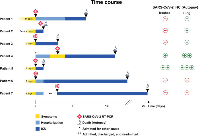
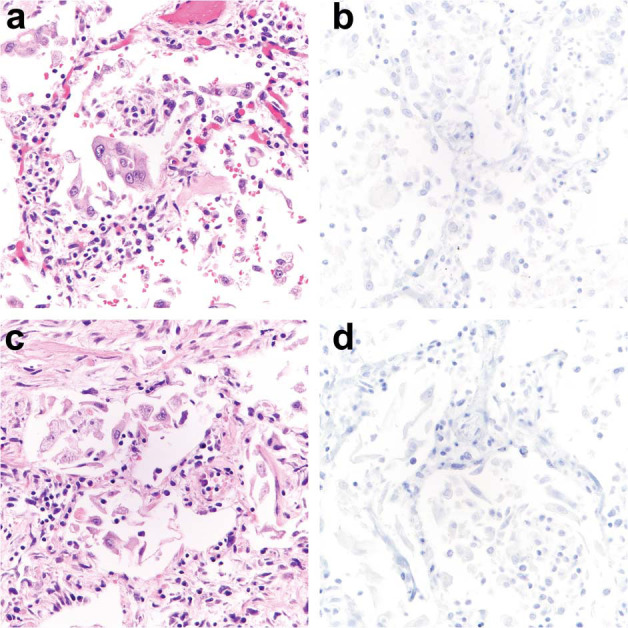
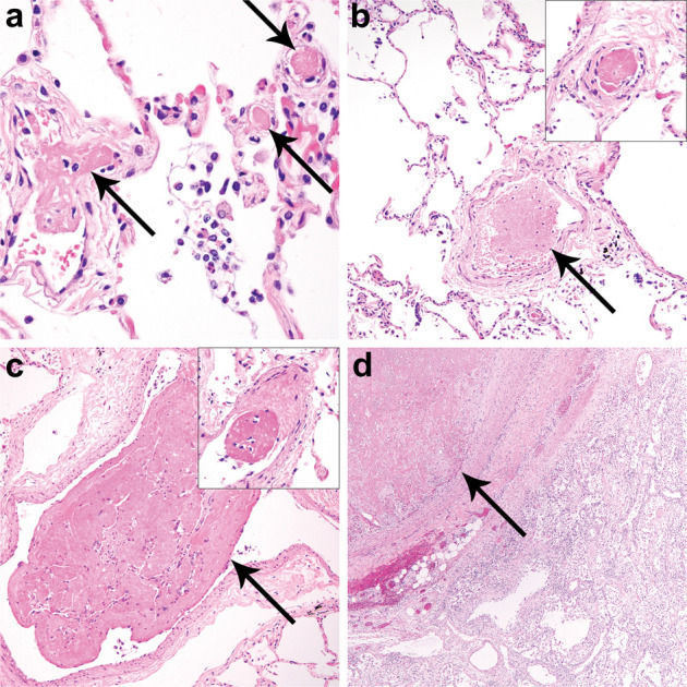
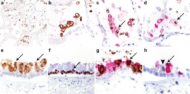
Coronavirus disease 2019 (COVID-19) caused by SARS-CoV-2 has led to a global public health crisis. In elderly individuals and those with comorbidities, COVID-19 is associated with high mortality, frequently caused by acute respiratory distress syndrome. We examine in situ expression of SARS-CoV-2 in airways and lung obtained at autopsy of individuals with confirmed COVID-19 infection. Seven autopsy cases (male, N = 5; female, N = 2) with reverse transcriptase-polymerase chain reaction (RT-PCR)-confirmed SARS-CoV-2 infection and a median age of 66 years (range, 50-77 years) were evaluated using a rabbit polyclonal antibody against SARS Nucleocapsid protein in correlation with clinical parameters. The median time from symptom onset to death was 9 days (range, 6-31 days), from hospitalization 7 days (range, 1-21 days), from positive RT-PCR 7 days (range, 0-18 days), and from intensive care unit admission defining onset of respiratory failure 3 days (range, 1-18 days). Chest imaging identified diffuse airspace disease in all patients corresponding to acute and (N = 5) or organizing (N = 2) diffuse alveolar damage (DAD) on histologic examination. Among five patients with acute-phase DAD (≤7 days from onset of respiratory failure), SARS-CoV-2 was detected in pulmonary pneumocytes and ciliated airway cells (N = 5), and in upper airway epithelium (N = 2). In two patients with organizing DAD (>14 days from onset of respiratory failure), no virus was detected in lungs or airways. No endothelial cell infection was observed. The findings suggest that SARS-CoV-2 infection of epithelial cells in lungs and airways of patients with COVID-19 who developed respiratory failure can be detected during the acute phase of lung injury and is absent in the organizing phase.


Figures

Fig. 1
Fig. 1
Clinical time course in seven patients with COVID-19 (SARS-CoV-2 infection). The schematic illustrates symptom duration, hospitalization, admission to intensive care unit (ICU), detection of SARS-CoV-2 by reverse transcriptase-polymerase chain reaction (RT-PCR), and death followed by autopsy examination. *Patient 4 was admitted for scheduled B-lymphoblastic leukemia treatment and developed symptoms 2 days after admission. **Patient 7 was hospitalized with symptoms, discharged, and readmitted 3 days later. Results of SARS immunohistochemistry (IHC) on autopsy trachea and lung tissue are shown on the right; “+” indicates positive staining in <5 cells per 4 mm2, “++” indicates positive staining in 5–50 cells per 4 mm2, and “+++” positive staining in >50 cells per 4 mm2.
Fig. 2
Fig. 2
Results of radiologic imaging and autopsy findings in patient 5 with COVID-19 (SARS-CoV-2 infection). Chest x-ray shows extensive interstitial and airspace opacities and cardiomegaly (a). CT imaging obtained the same day (b) demonstrates bilateral diffuse ground-glass opacities with subpleural sparing and linear band-like opacities (b, arrows). Cross section of the left lung at autopsy (c) reveals patchy consolidation involving the upper and lower lobes representing SARS-CoV-2 pneumonia (c, arrow). Histologic examination of the lungs identified acute-phase diffuse alveolar damage (d) with extensive hyaline membrane formation (d, arrow) desquamation of pulmonary epithelial cells into the alveolar spaces, and inflammatory infiltrates (d, inset).
Fig. 3
Fig. 3
Radiologic imaging in six patients with COVID-19 (SARS-CoV-2 infection). Chest x-rays in patients 1 (a), 2 (b), 3 (c), 4 (d), 6 (e), and 7 (f) showed extensive interstitial and airspace opacities consistent with COVID-19.
Fig. 4
Fig. 4
Histologic lung findings at autopsy and SARS immunohistochemical staining in lungs from patients 1–4 with COVID-19 (SARS-CoV-2 infection). Histologic examination of the lungs from patients 1 (a, b), 2 (c, d), 3 (e, f), and 4 (g, h) identified acute-phase diffuse alveolar damage (DAD) with extensive hyaline membrane formation (a, c, e, f, arrows). SARS immunohistochemistry (IHC) showed <5 cells with strongly positive staining per 4 mm2 in patients 1 (b, arrow, inset), 2 (d, inset), and 3 (f, inset). In patient 4, 5–50 cells with strongly positive staining were identified per 4 mm2 (h, inset).
Fig. 5
Fig. 5
Histologic lung findings at autopsy and SARS immunohistochemical staining in lungs from patients 6 and 7 with COVID-19 (SARS-CoV-2 infection). Histologic examination of the lungs from patients 6 (a, b), and 7 (c, d) showed organizing diffuse alveolar damage (DAD) (a, c) without prominent hyaline membrane formation that would correspond to acute-phase DAD. SARS immunohistochemistry (IHC) was entirely negative in lung sections from patient 6 (b) and 7 (d).
Fig. 6
Fig. 6
Histologic evidence of coagulopathy in patients with COVID-19 (SARS-CoV-2 infection). Histologic examination of the lungs identified multiple thromboemboli in small to intermediate sized intrapulmonary arteries and post-capillary venules of patients 2 (a, arrows), 3 (b, arrow, inset), and 5 (c, arrow, inset) and a large thromboembolus in patient 7 (d, arrow).
Fig. 7
Fig. 7
SARS immunohistochemical staining in lung and airways from patient 5 with COVID-19 (SARS-CoV-2 infection). SARS immunohistochemistry (IHC) on lung sections shows diffuse strong SARS positivity in pneumocytes in >50 cells per 4 mm2 (a, b). TTF-1/SARS double labeling (c) demonstrates co-localization of TTF-1 (brown reaction product) and SARS (red reaction product) in pneumocytes (c, arrow). PU.1/SARS double labeling demonstrates expression of PU.1 (brown reaction product) in macrophages, most of which did not stain for SARS (red reaction product), and SARS mostly in pneumocytes negative for PU.1 (d). Weak staining for SARS was identified only in scattered macrophages (d, arrow). SARS IHC on cross sections of trachea (e) demonstrates positive staining predominantly in ciliated cells (e, arrows). P63/SARS double labeling demonstrates nuclear expression of p63 in basal cells (brown reaction product) (f, arrow) which did not stain for SARS (red reaction product). MUC5AC/SARS double labeling shows cytoplasmic expression of MUC5AC in secretory cells (brown reaction product) (g, arrow) most of which did not stain for SARS (red reaction product). FOXJ1 highlighted uninfected ciliated cells (h, arrowhead) but was absent in SARS-expressing cells (h, arrow).

Similar articles

Cited by

References

    1. Coronaviridae Study Group of the International Committee on Taxonomy of V. The species severe acute respiratory syndrome-related coronavirus: classifying 2019-nCoV and naming it SARS-CoV-2. Nat Microbiol. 2020;5:536–544. doi: 10.1038/s41564-020-0695-z. - DOI - PMC - PubMed
    1. Guan WJ, Ni ZY, Hu Y, Liang WH, Ou CQ, He JX, et al. Clinical characteristics of coronavirus disease 2019 in China. N Engl J Med. 2020;18:1708–1720. doi: 10.1056/NEJMoa2002032. - DOI - PMC - PubMed
    1. Yang X, Yu Y, Xu J, Shu H, Xia J, Liu H, et al. Clinical course and outcomes of critically ill patients with SARS-CoV-2 pneumonia in Wuhan, China: a single-centered, retrospective, observational study. Lancet Respir Med. 2020;8:475–481. doi: 10.1016/S2213-2600(20)30079-5. - DOI - PMC - PubMed
    1. Goyal P, Choi JJ, Pinheiro LC, Schenck EJ, Chen R, Jabri A, et al. Clinical characteristics of Covid-19 in New York City. N Engl J Med. 2020;382:2372–2374. doi: 10.1056/NEJMc2010419. - DOI - PMC - PubMed
    1. Tian S, Hu W, Niu L, Liu H, Xu H, Xiao SY. Pulmonary pathology of early-phase 2019 novel coronavirus (COVID-19) pneumonia in two patients with lung cancer. J Thorac Oncol. 2020;5:700–704. doi: 10.1016/j.jtho.2020.02.010. - DOI - PMC - PubMed