. 2020 Jun 19;6(1):143.
doi: 10.1186/s40792-020-00905-x.

Invasive solid papillary carcinoma with neuroendocrine differentiation of the breast: a case report and literature review

Affiliations

Invasive solid papillary carcinoma with neuroendocrine differentiation of the breast: a case report and literature review

Xue Lin et al. Surg Case Rep. .

Abstract

Background: Solid papillary carcinoma (SPC) of the breast is a rare breast cancer that accounts for less than 1% of all breast cancers. The optimal clinical management of SPC remains controversial. Here, we report a case of invasive SPC with neuroendocrine differentiation in addition to review of the current literature.

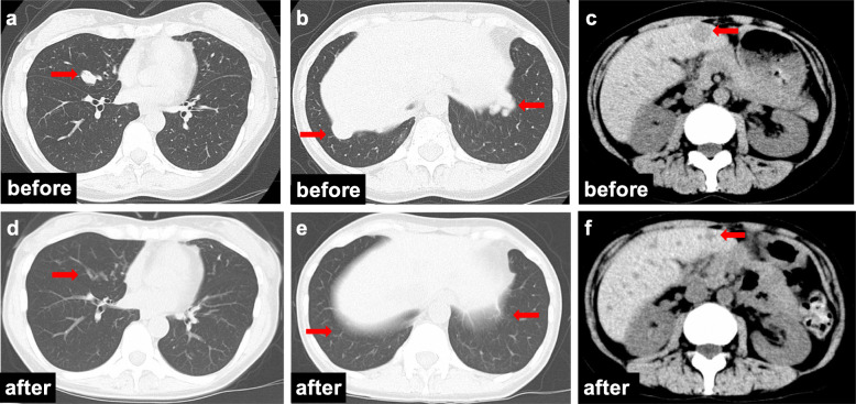
Case presentation: A premenopausal 46-year-old female presented with a mass in her left breast that tended to increase in size over a 10-month period. Mammography and ultrasonography revealed a mass in the left upper-inner quadrant. The resulting images suggested a category 3 breast tumor according to the Breast Imaging Reporting and Data System (BI-RADS). A core needle biopsy (CNB) was performed, and the pathological findings showed a solid papillary pattern and atypical cells suggestive of noninvasive SPC. After a left partial mastectomy and sentinel lymph node biopsy (SLNB), the specimens were sent for histopathological analysis for further investigation. Postoperative pathological findings suggested invasive SPC. Whole-breast radiation therapy and adjuvant hormonal therapy were performed as postoperative treatments. Three years after surgery, multiple lung metastases were detected, and the patient was treated with a gonadotropin-releasing hormone agonist and an aromatase inhibitor. Five months later, multiple liver metastases and bone metastases appeared, and oral 5-fluorouracil was chosen for the subsequent treatment. The patient has been treated for 5 years to date, and she is continuing to take oral 5-fluorouracil and is alive without any further disease progression.

Conclusions: We report a rare case of premenopausal invasive SPC with multiple metastases. Further study is needed to clarify the molecular characteristics and clinical behaviors of SPC and to explore the optimal treatment strategy.

Keywords: Breast; Invasive solid papillary carcinoma; Metastasis; Neuroendocrine differentiation.


Conflict of interest statement

The authors declare that they have no competing interests.

Figures

Fig. 1
Fig. 1
Imaging findings. Mammogram showing a round, high-density mass in the left MLO-M area (a) and CC-I area (c). b, d Enlargements of the areas are indicated by red arrows. US shows a regular, circumscribed, hypoechoic, homogenous lesion with abundant blood flow in the A area of the left breast (e). Dynamic MRI demonstrates an ill-demarcated mass that measures 8 mm × 7 mm in the A area of the left breast (f) without daughter lesions or enlarged lymph nodes
Fig. 2
Fig. 2
Core needle biopsy histological findings of the tumor (a) and (b) HE-stained sections at × 4 and × 20 magnification. a Low-power view illustrates a solid papillary carcinoma. b High-power view demonstrates the rosette structure of a solid papillary growth pattern and a network of thick fibrovascular cores among proliferating atypical malignant cells. c Positive immunohistochemical staining findings for the estrogen receptor. Positive immunohistochemical staining findings for the following markers demonstrate the neuroendocrine component of the tumor: d neuron-specific enolase, e chromogranin, and f synaptophysin. g Negative immunohistochemical staining for the myoepithelial marker p63 (a: × 4, bg: × 20)
Fig. 3
Fig. 3
Postoperative histological findings of the tumor. a Postoperative surgical specimen of the left breast tissue with a visible tumor (red arrow). b HE-stained section of the tumor at × 4 magnification. c HE-stained section of the tumor at × 20 magnification. The HE-stained sections illustrate a clear infiltration that suggests an invasive solid papillary carcinoma. The invasive components that spread out from the solid structure are visible (red arrow, b, c)
Fig. 4
Fig. 4
Whole-body CT before and after chemotherapy. Lung metastatic nodules (a, b) and liver tumors (c) were detected. According to treatment with oral 5-FU, the lung metastatic tumors completely disappeared (d, e), and the liver metastases remarkably regressed (f)

References

    1. Rakha EA, Ahmed MA, Ellis IO. Papillary carcinoma of the breast: diagnostic agreement and management implications. Histopathology. 2016;69:862–870. doi: 10.1111/his.13009. - DOI - PubMed
    1. Saremian J, Rosa M. Solid papillary carcinoma of the breast: a pathologically and clinically distinct breast tumor. Arch Pathol Lab Med. 2012;136:1308–1311. doi: 10.5858/arpa.2011-0227-RS. - DOI - PubMed
    1. Maluf HM, Koerner FC. Solid papillary carcinoma of the breast. A form of intraductal carcinoma with endocrine differentiation frequently associated with mucinous carcinoma. Am J Surg Pathol. 1995;19:1237–1244. doi: 10.1097/00000478-199511000-00003. - DOI - PubMed
    1. Otsuki Y, Yamada M, Shimizu S, Suwa K, Yoshida M, Tanioka F, et al. Solid-papillary carcinoma of the breast: clinicopathological study of 20 cases. Pathol Int. 2007;57:421–429. doi: 10.1111/j.1440-1827.2007.02118.x. - DOI - PubMed
    1. Yamada M, Otsuki Y, Shimizu S, Tanioka F, Ogawa H, Kobayashi H. Cytological study of 20 cases of solid-papillary carcinoma of the breast. Diagn Cytopathol. 2007;35:417–422. doi: 10.1002/dc.20668. - DOI - PubMed

LinkOut - more resources