. 2021 Feb;78(3):1085-1100.
doi: 10.1007/s00018-020-03570-3. Epub 2020 Jun 19.

KDEL receptor is a cell surface receptor that cycles between the plasma membrane and the Golgi via clathrin-mediated transport carriers

Affiliations

KDEL receptor is a cell surface receptor that cycles between the plasma membrane and the Golgi via clathrin-mediated transport carriers

Jie Jia et al. Cell Mol Life Sci. 2021 Feb.

Abstract

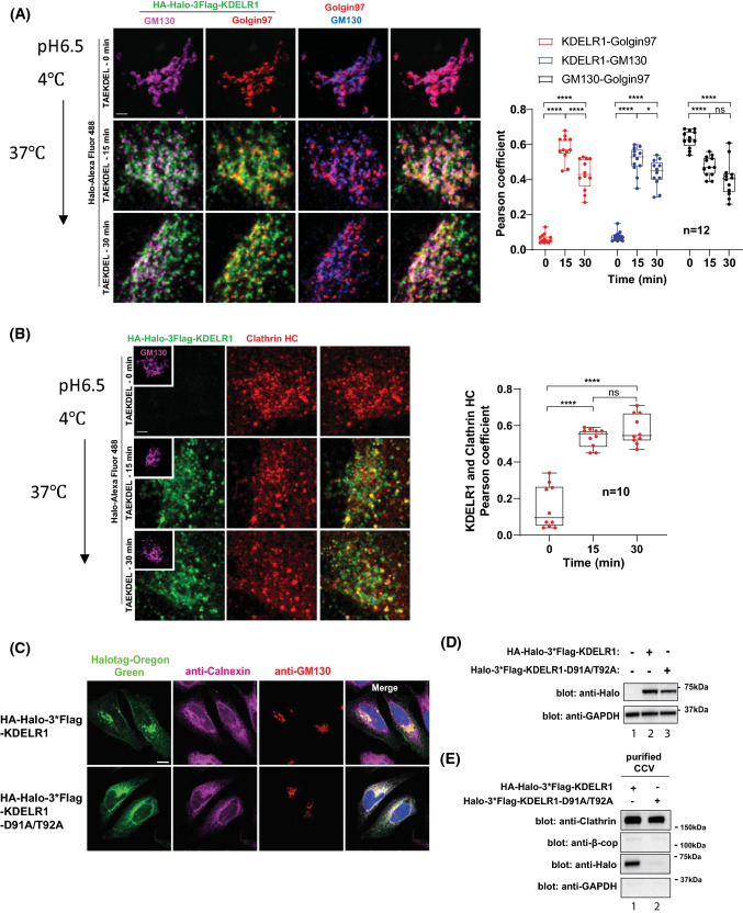
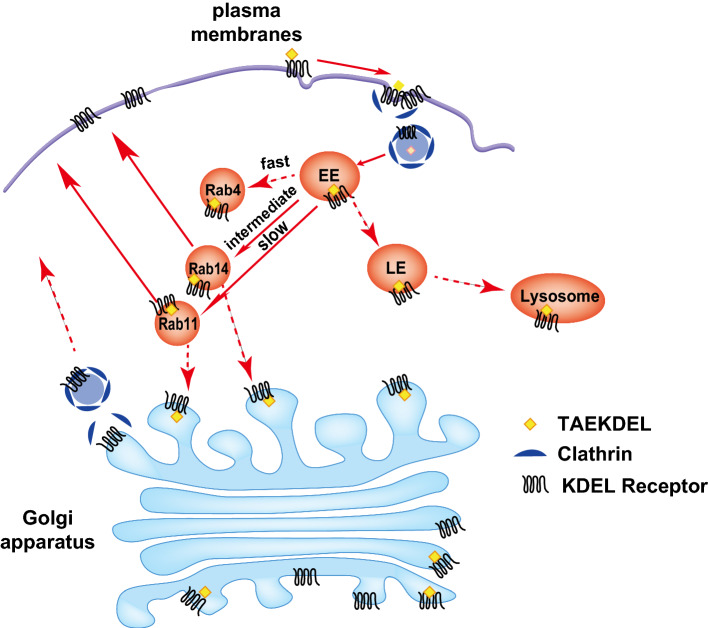
KDEL receptor cycles between the ER and the Golgi to retrieve ER-resident chaperones that get leaked to the secretory pathway during protein export from the ER. Recent studies have shown that a fraction of KDEL receptor may reside in the plasma membrane and function as a putative cell surface receptor. However, the trafficking itinerary and mechanism of cell surface expressed KDEL receptor remains largely unknown. In this study, we used N-terminally Halo-tagged KDEL receptor to investigate its endocytosis from the plasma membrane and trafficking itinerary of the endocytosed receptor through the endolysosomal compartments. Our results indicate that surface-expressed KDEL receptor undergoes highly complex recycling pathways via the Golgi and peri-nuclear recycling endosomes that are positive for Rab11 and Rab14, respectively. Unexpectedly, KDEL receptor appears to preferentially utilize clathrin-mediated endocytic pathway as well as clathrin-dependent transport carriers for export from the trans-Golgi network. Taken together, we suggest that KDEL receptor may be a bona fide cell surface receptor with a complex, yet well-defined trafficking itinerary through the endolysosomal compartments.

Keywords: Caveolae; Clathrin; EEA1; Early endosomes; Golgi; KDEL receptor; Late endosomes; Lysosome; MANF; Membrane trafficking; Rab11; Rab14; Recycling endosomes.


Conflict of interest statement

The authors declare no competing interests.

Figures

Fig. 1
Fig. 1
Surface-expressed wild type KDELR1, but not the H12A mutant receptor, binds TAEKDEL peptide. a Comparison of cell surface expression level of three isoforms of KDELR. MCF7 cells were transiently transfected with KDELR1-mCherry, KDELR2-mCherry or KDELR3-mCherry for 18 h. Cell surface expression of KDELR-mCherry was probed by cell surface biotinylation protocol, as described in the methods. b Schematic representation of the integration strategy to generate C-terminally 3xFlag-mCherry/Halo-tagged proteins expressed from the endogenous KDELR1 locus. A double-strand break was created at the last exon of KDELR1 gene by Cas9 RNP. Gene knock-in was mediated by a plasmid DNA donor template that contains 3xFlag-mCherry/Halo sequence, neomycin resistant gene and homology arms. c Cell surface biotinylation of endogenously tagged KDELR1. Cell surface proteins were biotinylated by treatment with (+) or without (−) Sulfo-NHS-LC-Biotin in KDELR1-knock-in (C-terminal 3xFlag-mCherry endogenously tagged) HeLa cells. Biotinylated proteins were isolated by streptavidin-agarose and subjected to western blot analysis using the indicated antibodies. Whole cell lysates (input) served as control to determine the total amount of KDELR1, while GM130 served as a cytosolic marker protein, which showed no biotinylation and EGFR served as a plasma membrane marker protein, respectively. Membrane fraction (surface) illustrates the total fraction of proteins at the cell surface. d Schematic representation of Halo-tagged KDEL receptor. e Expression of Halo-KDELR and Halo-KDELR H12A in MCF7 KDELR KD cells was detected by immunoblotting. f Comparison of the expression level of over-expressed HA-Halo-3xFlag-tagged KDELR1 in HeLa wt, MCF7 wt and MCF KDELR1 KD cells with that of endogenously 3xFlag-Halo-tagged KDELR1 in HeLa cells by immunoblotting. g MCF7 KDELR KD cells were transfected with either Halo-KDELR or Halo-KDELR H12A for 18 h. Living cells were then stained with HaloTag Alexa Fluor 488 and HaloTag TMR ligands at 4 °C for 30 min, followed by fixation and DAPI staining. The subcellular localization of Halo-KDELR or Halo-KDELR H12A was observed using Zeiss LSM880. hi Halo-KDELR (h) or Halo-KDELR H12A (i) expressed in MCF7 KDELR KD cells was incubated with HaloTag Alexa Fluor 488 ligand and 50 μM TMR-TAEKDEL peptide in DMEM (pH 6.5) supplemented with 10% FBS at 4 °C for 30 min. Cells were then washed 3 times with PBS and incubated at 37 °C for 0, 15, and 30 min, followed by fixation. Confocal images were acquired using Zeiss LSM880. Scale bar: 5 µm
Fig. 2
Fig. 2
Surface-expressed KDELR undergo clathrin-mediated endocytosis. a Live-cell imaging of clathrin-dependent endocytosis of KDELR1. HeLa cells were co-transfected with Halo-KDELR1 and mCherry-CLC (clathrin light chain) for 18 h. Living cells were preincubated with HaloTag Alexa Fluor 488 ligand at 4 °C for 30 min. Cells were then washed with PBS and observed using Zeiss Airyscan confocal microscope at 37 °C for 30 min in live cell imaging medium (pH 6.5), supplemented with 1% FBS and 50 μM TAEKDEL or TAEAAAA peptides. Arrowheads indicate punctae containing both Halo-KDELR and mCherry-CLC proteins. Scale bars: 10 µm. Insets show a magnified view of the boxed area. Line profiles through regions of interest were analyzed by Fiji. b TIRF imaging showed endocytosis of KDELR1 was dependent of clathrin rather than caveolin. MCF7 cells overexpressing Halo-KDELR were incubated with HaloTag Alexa Fluor 488 ligand and 50 μM TAEKDEL peptide in DEMEM (pH 6.5) supplemented with 10% FBS at 4 °C for 30 min. Cells were then washed 3 times with ice-cold PBS and incubated at 37 °C for 0 and 30 min, followed by fixation and staining by anti-clathrin or anti-caveolin antibodies, respectively. Images were acquired using Nikon TIRF microscope. Box graphs summarize the co-localization of Halo-KDELR1 with clathrin or caveolin, respectively (n = 10 cells). Statistical analysis was performed using two-way ANOVA with a Tukey's post-hoc test for multiple comparisons. Insets show a magnified view of the boxed area. Line profiles through regions of interest were analyzed by Fiji. Scale bars: 10 µm. *** p < 0.001, **** p < 0.0001, ns not significant
Fig. 3
Fig. 3
Surface-expressed KDELR is transported to the early endosomes upon endocytosis. Co-localization of endocytosed Halo-KDELR and intracellular organelles was determined using confocal microscopy. a MCF7 KDELR KD cells were transfected with either Halo-KDELR or Halo-KDELR H12A with mCherry-tagged Rab7 (d). After 18 h, cells were treated with DMSO (a and d) or MitoTracker (b) or LysoTracker (c) at 37 °C for 30 min and then incubated with HaloTag Alexa Fluor 488 ligand and TAEKDEL peptide in DMEM (pH 6.5) with 10% FBS at 4 °C for 30 min. Cells were then washed with PBS and transferred to 37 °C for 0, 15, or 30 min, prior to fixation and staining with anti-EEA1 antibody (a). The colocalization between HA-Halo-KDELR (WT or H12A) and various organelles was quantified using Pearson’s coefficient and summarized by line graph (n = 20 cells). Statistical analysis was performed using two-way ANOVA with a Tukey's post-hoc test for multiple comparisons. Scale bars: 5 µm. * p < 0.05, *** p < 0.001, **** p < 0.0001, #p < 0.0001, ns not significant
Fig. 4
Fig. 4
RTDL motif of MANF can also accelerate trafficking of endocytosed KDELR1 to the early endosomes. MCF7 cells were transfected with HA-Halo-KDELR1 for 18 h and incubated with HaloTag Alexa Fluo 488 ligand and 50 μM TAEKDEL (a), 50 μM ASARTDL (b) or DMSO (c) in DMEM (pH 6.5) with 10% FBS at 4 °C for 30 min. Cells were then washed with PBS and transferred to 37 °C for indicated times, prior to fixation and staining with anti-EEA1 antibody. Insets and line graphs show the colocalization details of HA-Halo-KDELR and EEA1. Pearson’s coefficient was used to quantify the colocalization between HA-Halo-KDELR and EEA1. Statistical analysis was performed using two-way ANOVA with a Tukey's post-hoc test for multiple comparisons. Scale bar: 5 µm. ** p < 0.01, **** p < 0.0001, #p < 0.0001, ##p < 0.01, ###p < 0.001
Fig. 5
Fig. 5
A fraction of endocytosed KDELR enters the Golgi and is recycled to the endosomal compartments or the PM via clathrin-mediated transport carriers. a Co-localization of endocytosed KDELR with the Golgi apparatus was investigated by Leica confocal microscope in HeLa cells treated with 50 μM TAEKDEL peptide for indicated times and costained with GM130 (cis-Golgi) and Golgin97 (trans-Golgi). n = 12, Statistical analysis was performed using one-way ANOVA with a Tukey's post-hoc test. Scale bars: 2 µm. b Co-localization of endocytosed KDELR with endogenous clathrin in HeLa cells treated with 50 μM TAEKDEL peptide for indicated times was examined by Leica confocal microscopy using antibody against clathrin heavy chain. n = 10, Statistical analysis was performed using one-way ANOVA with a Tukey's post-hoc test. Scale bars: 2 µm. c HeLa cells transfected with HA-Halo-KDELR1 or Halo-KDELR1 D91A/T92A were stained with membrane permeable-Halotag Oregon Green, anti-calnexin antibody, anti-GM130 antibody, and DAPI. Confocal images were obtained by Zeiss LSM880. Scale bar: 5 µm. d The protein expression of KDELR1 WT and D91A/T92A mutant was analyzed by immunoblotting of HeLa cells transfected with HA-Halo-KDELR1 or HA-Halo-KDELR1 D91A/T92A. Endogenous GAPDH was detected as a control. e HeLa cells were transfected with HA-Halo-KDELR1 or HA-Halo-KDELR1 D91A/T92A for 18 h and then collected to purify clathrin-coated vesicles (CCV) according to a published protocol (23). CCVs were further analyzed by SDS-PAGE and western blotting with specific antibodies against Halo, clathrin heavy chain, β-COP and GAPDH. HA-Halo-KDELR1, but not Halo-KDELR1 D91A/T92A, was detected in CCVs. * p < 0.05, **** p < 0.0001, ns not significant
Fig. 6
Fig. 6
Endocytosed KDELR is recycled via peri-nuclear recycling pathways. Colocalization of endocytosed Halo-KDELR and mCherry-tagged Rab11 (a), Rab14 (b) and Rab4 (c) was determined using confocal microscopy. MCF7 KDELR KD cells transfected with Halo-KDELR or Halo-KDELR H12A and mCherry-tagged Rab 11 (a), Rab14 (b), or Rab4 (c) were incubated with HaloTag Alexa Fluor 488 ligand and TAEKDEL peptide in DMEM (pH 6.5) with 10% FBS at 4 °C for 30 min, followed by washing with PBS and incubated at 37 °C for 0, 15, or 30 min. Cells were then fixed and observed using Zeiss LSM880. Line graphs indicate the colocalization of Halo-KDELR (WT or H12A) and Rab proteins (n = 20 cells). Statistical analysis was performed using two-way ANOVA with a Tukey's post-hoc test for multiple comparisons. Scale bars: 10 µm. * p < 0.05, ** p < 0.01, **** p < 0.0001, #p < 0.0001, ns not significant
Fig. 7
Fig. 7
Schematic illustration that depicts intracellular itinerary of surface-expressed KDELR. Our results indicate that surface-expressed KDELR undergoes clathrin-mediated endocytosis, which could be further accelerated upon binding of KDEL ligand to the receptor. Endocytosed KDELR then travels to early endosomes (EE), where initial sorting of the endocytosed receptor seems to occur. From the EE, bulk of endocytosed KDELR seems to undergo recycling through peri-nuclear recycling pathways via Rab14- and Rab11-positive recycling endosomes. A fraction of endocytosed KDELR travels to the Golgi and is rapidly recycled to the endosomal compartments or the PM via clathrin-mediated transport carriers at the TGN. It is currently unknown what fraction of endocytosed KDELR in the Rab14- and Rab11-positive endosomes may transit through the Golgi, prior to recycling to the cell surface

Similar articles

Cited by

References

    1. Munro S, Pelham HR. A C-terminal signal prevents secretion of luminal ER proteins. Cell. 1987;48:899–907. doi: 10.1016/0092-8674(87)90086-9. - DOI - PubMed
    1. Lewis MJ, Pelham HR. A human homologue of the yeast HDEL receptor. Nature. 1990;348:162–163. doi: 10.1038/348162a0. - DOI - PubMed
    1. Lewis MJ, Sweet DJ, Pelham HR. The ERD2 gene determines the specificity of the luminal ER protein retention system. Cell. 1990;61:1359–1363. doi: 10.1016/0092-8674(90)90699-F. - DOI - PubMed
    1. Pulvirenti T, et al. A traffic-activated Golgi-based signalling circuit coordinates the secretory pathway. Nat Cell Biol. 2008;10:912–922. doi: 10.1038/ncb1751. - DOI - PubMed
    1. Capitani M, Sallese M. The KDEL receptor: new functions for an old protein. FEBS Lett. 2009;583:3863–3871. doi: 10.1016/j.febslet.2009.10.053. - DOI - PubMed

MeSH terms

LinkOut - more resources