Imaging in Vasculitis
- PMID: 32562073
- PMCID: PMC7305069
- DOI: 10.1007/s11926-020-00915-6
Imaging in Vasculitis
Abstract
Purpose of review: Vasculitides are characterized by mostly autoimmunologically induced inflammatory processes of vascular structures. They have various clinical and radiologic appearances. Early diagnosis and reliable monitoring are indispensable for adequate therapy to prevent potentially serious complications. Imaging, in addition to laboratory tests and physical examination, constitutes a key component in assessing disease extent and activity. This review presents current standards and some typical findings in the context of imaging in vasculitis with particular attention to large vessel vasculitides.
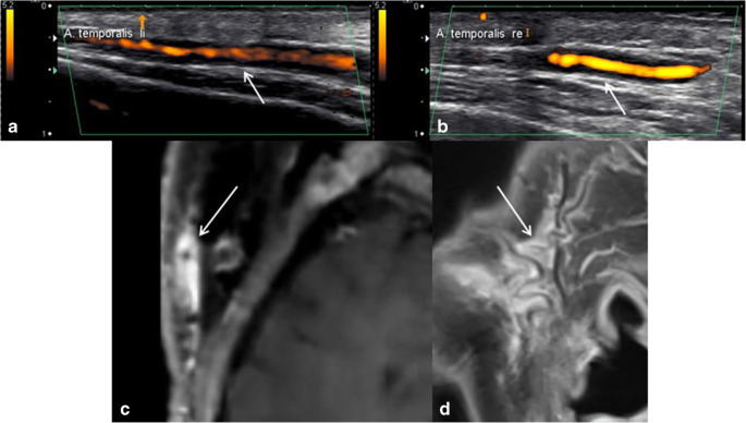
Recent findings: Recently, imaging has gained importance in the management of vasculitis, especially regarding large vessel vasculitides (LVV). Recently, EULAR (European League Against Rheumatism) has launched its recommendations concerning the diagnosis of LVVs. Imaging is recommended as the preferred complement to clinical examination. Color-coded duplex sonography is considered the first choice imaging test in suspected giant cell arteritis, and magnetic resonance imaging is considered the first choice in suspected Takayasu's arteritis. Due to diversity of clinical and radiologic presentations, diagnosis and therapy monitoring of vasculitides may constitute a challenge. As a result of ongoing technological progress, a variety of non-invasive imaging modalities now play an elemental role in the interdisciplinary management of vasculitic diseases.
Keywords: EULAR guidelines; Giant cell arteritis (GCA); Imaging; Large vessel vasculitides (LVV); Magnetic resonance imaging (MRI); Vasculitis.
Conflict of interest statement
The authors declare that they have no conflict of interest.
Figures


Similar articles
-
Imaging in Large Vessel Vasculitides.Rofo. 2019 Dec;191(12):1083-1090. doi: 10.1055/a-0896-2833. Epub 2019 May 16. Rofo. 2019. PMID: 31096287 Review. English.
-
EULAR recommendations for the use of imaging in large vessel vasculitis in clinical practice.Ann Rheum Dis. 2018 May;77(5):636-643. doi: 10.1136/annrheumdis-2017-212649. Epub 2018 Jan 22. Ann Rheum Dis. 2018. PMID: 29358285
-
Ultrasound imaging in the diagnosis of large vessel vasculitis.Vasa. 2017 Jul;46(4):241-253. doi: 10.1024/0301-1526/a000625. Epub 2017 Mar 23. Vasa. 2017. PMID: 28332442 Review.
-
Imaging for Diagnosis, Monitoring, and Outcome Prediction of Large Vessel Vasculitides.Curr Rheumatol Rep. 2020 Sep 21;22(11):76. doi: 10.1007/s11926-020-00955-y. Curr Rheumatol Rep. 2020. PMID: 32959107 Free PMC article. Review.
-
EULAR recommendations for the use of imaging in large vessel vasculitis in clinical practice summary.Radiol Med. 2019 Oct;124(10):965-972. doi: 10.1007/s11547-019-01058-0. Epub 2019 Jun 28. Radiol Med. 2019. PMID: 31254221 Review.
Cited by
-
Granulomatosis with polyangiitis: clinical characteristics and updates in diagnosis.Front Med (Lausanne). 2024 Aug 27;11:1369233. doi: 10.3389/fmed.2024.1369233. eCollection 2024. Front Med (Lausanne). 2024. PMID: 39257888 Free PMC article. Review.
-
Migrant cavitation as primary involvement in a particular case of granulomatosis with polyangiitis.Radiol Case Rep. 2022 Jul 28;17(10):3535-3538. doi: 10.1016/j.radcr.2022.06.074. eCollection 2022 Oct. Radiol Case Rep. 2022. PMID: 35923342 Free PMC article.
-
Ultrasound imaging of the dorsalis pedis artery as an early indicator of the precursory changes for rheumatoid vasculitis: A case series.Australas J Ultrasound Med. 2023 Dec 17;27(1):42-48. doi: 10.1002/ajum.12373. eCollection 2024 Feb. Australas J Ultrasound Med. 2023. PMID: 38434542 Free PMC article.
-
Angiography Revealing a Possible Paraneoplastic Vasculitis: A Case Report.Cureus. 2024 Jun 26;16(6):e63229. doi: 10.7759/cureus.63229. eCollection 2024 Jun. Cureus. 2024. PMID: 39070390 Free PMC article.
-
Pediatric cardiothoracic vasculitis: multimodality imaging review.Pediatr Radiol. 2022 Sep;52(10):1895-1909. doi: 10.1007/s00247-022-05431-6. Epub 2022 Jul 6. Pediatr Radiol. 2022. PMID: 35790558 Free PMC article. Review.
References
-
- Muhle C, Reinhold-Keller E, Richter C, Duncker G, Beigel A, Brinkmann G, et al. MRI of the nasal cavity, the paranasal sinuses and orbits in Wegener’s granulomatosis. Eur Radiol. 1997;7:566–70. - PubMed
Publication types
MeSH terms
LinkOut - more resources
Full Text Sources
Medical
Research Materials