. 2020 Sep;59(9):1052-1063.
doi: 10.1002/mc.23236. Epub 2020 Jun 20.

Loss of prdm1a accelerates melanoma onset and progression

Affiliations

Loss of prdm1a accelerates melanoma onset and progression

Ritsuko Iwanaga et al. Mol Carcinog. 2020 Sep.

Abstract

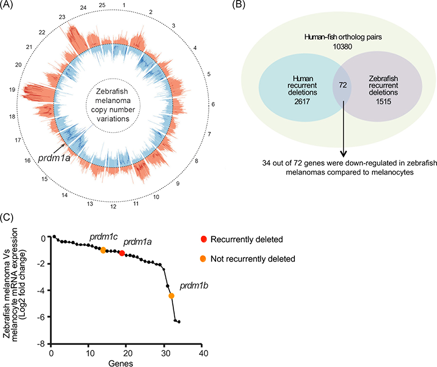
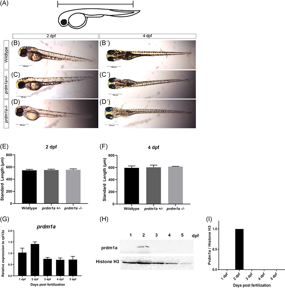
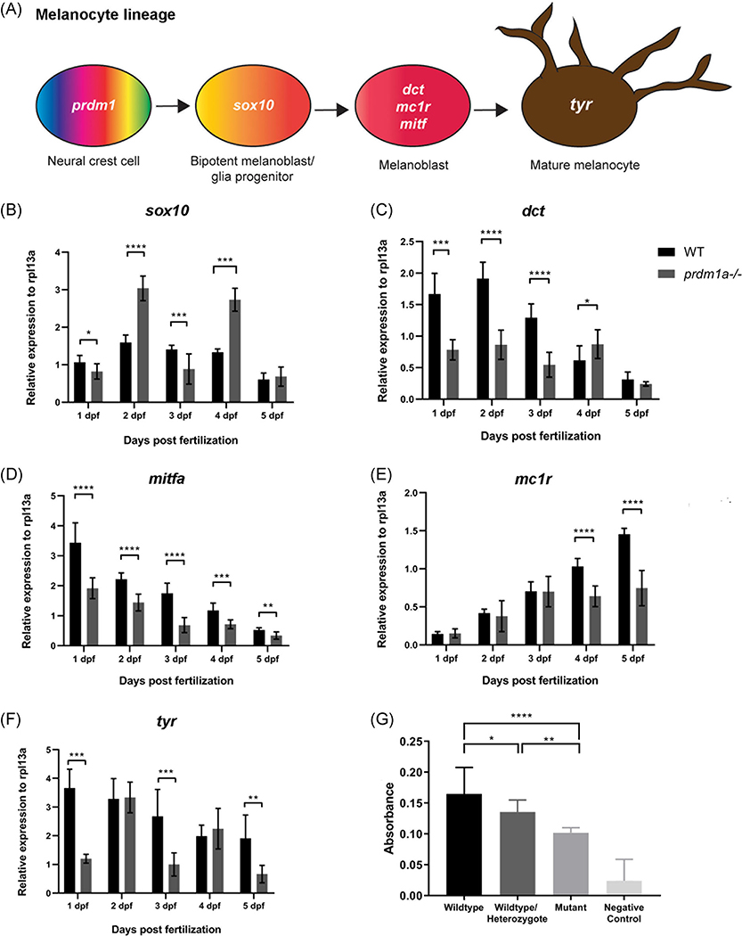
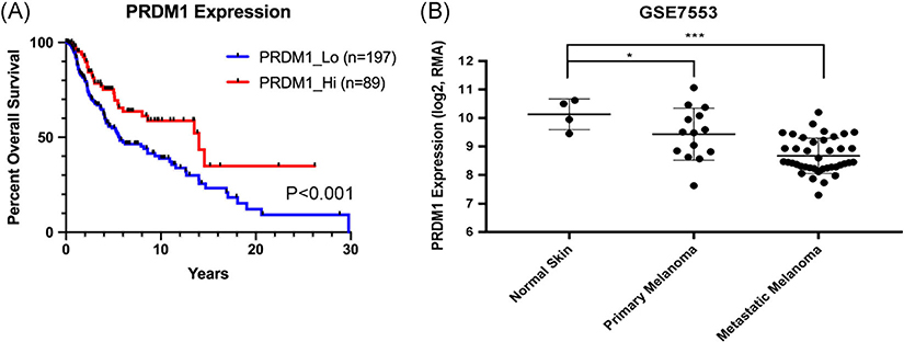
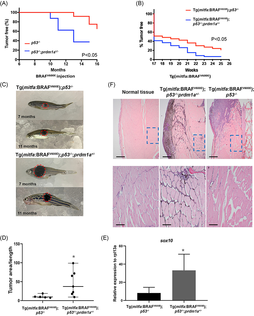
Melanoma is an aggressive, deadly skin cancer derived from melanocytes, a neural crest cell derivative. Melanoma cells mirror the developmental program of neural crest cells in that they exhibit the same gene expression patterns and utilize similar cellular mechanisms, including increased cell proliferation, epithelial-mesenchymal transition, and migration. Here we studied the role of neural crest regulator PRDM1 in melanoma onset and progression. In development, Prdm1a functions to promote neural crest progenitor fate, and in melanoma, we found that PRDM1 has reduced copy number and is recurrently deleted in both zebrafish and humans. When examining expression of neural crest and melanocyte development genes, we show that sox10 progenitor expression is high in prdm1a-/- mutants, while more differentiated melanocyte markers are reduced, suggesting that normally Prdm1a is required for differentiation. Data mining of human melanoma datasets indicates that high PRDM1 expression in human melanoma is correlated with better patient survival and decreased PRDM1 expression is common in metastatic tumors. When one copy of prdm1a is lost in the zebrafish melanoma model Tg(mitfa:BRAFV600E );p53-/- ;prdm1a+/- , melanoma onset occurs more quickly, and the tumors that form have a larger area with increased expression of sox10. These data demonstrate a novel role for PRDM1 as a tumor suppressor in melanoma.

Keywords: PRDM1; melanoma; neural crest cells; zebrafish.


Conflict of interest statement

CONFLICT OF INTERESTS

The authors declare that there are no conflict of interests.

Figures

FIGURE 1
FIGURE 1
PRDM1 is recurrently deleted in human and zebrafish melanoma. A, Circos plot displaying gene copy-number gains and losses in zebrafish melanomas as previously described. JISTIC G-scores are displayed as pale red shading (amplifications [minimum = 0; maximum = 1550]) and blue shading (deletions [minimum = 0; maximum = 250]). −log10-transformed JISTIC Q-values with a cutoff of 0.6 (corresponding to an untransformed Q-value of 0.25) are shown as bold red lines (amplifications [minimum = 0; maximum = 11]) and bold blue (deletions [minimum = 0; maximum = 11]). Dotted circles represent the –log10-transformed Q-value of 0 (center) and 11 (outer: amplification; inner: deletion). B, Venn diagram of orthologous genes significantly deleted in human and zebrafish melanomas from 10 380 human-zebrafish gene pairs (hypergeometric test, P value: 2.0E-16). C, Genes downregulated in zebrafish melanomas as compared with melanocytes (analyzed from previously obtained microarray dataset). prdm1a (red dot) and its paralogs prdm1b and prdm1c (orange dots) are indicated
FIGURE 2
FIGURE 2
prdm1aexpression during early development. A, Schematic showing how the standard length of the embryo was measured. Lateral images of wildtype (B), prdm1a+/− (C), and prdm1a−/− (D) at 2 dpf. Ventral images of wildtype (B’), prdm1a+/− (C’) and prdm1a−/− (D’) at 4 dpf. Quantification of standard length at 2 (E) and 4 (F) dpf. Statistical analysis was performed with a one-way ANOVA. G, RT-qPCR was performed on 20 pooled embryos at each time point (n = 3 replicate experiments) for prdm1a. H, Proteins were resolved by SDS-PAGE and Western blotting for prdm1a and total H3 was performed. I, Quantification of band intensity from the Western blot (H). Relative intensity was normalized to total histone H3 for each sample. ANOVA, analysis of variance; dpf, days postfertilization; SDS-PAGE, sodium dodecyl sulfate-polyacrylamide gel electrophoresis
FIGURE 3
FIGURE 3
prdm1a is required for melanocyte differentiation and pigment production. A, Melanocyte lineage from neural crest to mature melanocytes and their characteristic markers. Whole wildtype embryos were pooled for RNA isolation and protein extraction over developmental time. B-F, Whole wildtype and prdm1a−/− mutant embryos were pooled for RNA isolation over developmental time. Twenty whole embryos were pooled for each genotype and at each time point (n = 3 replicate experiments). RT-qPCR was performed for sox10 (B), dct (C), mitfa (D), mc1r (E), and tyr (F) over developmental time. Expression was compared using an unpaired, independent t test. J, 100 Embryos were pooled and incubated with L-DOPA for a tyrosinase functional assay. Pigment production was measured by spectrometry in whole wildtype, wildtype/heterozygote mix, and prdm1a/ mutant embryos. Statistical analysis was performed with a one-way ANOVA followed by a multiple comparisons test. Error bars represent mean ± SD. *P < .05, **P < .01, ***P < .001, ****P < .0001. ANOVA, analysis of variance; dpf, days postfertilization; L-DOPA, L-3,4-dihydroxyphenylalanine: SD, standard deviation
FIGURE 4
FIGURE 4
Low PRDM1 expression in humans is correlated with worse patient outcomes and metastatic melanoma. A, Overall survival of patients with metastatic melanoma compared to PRDM1 expression obtained from The Cancer Genome Atlas (TCGA). Statistical analysis was performed with a log-rank test. B, PRDM1 expression in normal skin, primary melanoma tumors, and metastatic melanoma tumors obtained from the Riker melanoma dataset. Statistical analysis was performed using a one-way ANOVA. Error bars represent mean ± SD. *P < .05, **P < .01, ***P < .001. ANOVA, analysis of variance; SD, standard deviation
FIGURE 5
FIGURE 5
Loss of prdm1a in zebrafish accelerates tumor onset. A, Tumor-free curve from p53−/− (n = 12) and p53−/−;prdm1a+/− (n = 8) zebrafish injected with BRAFV600E construct to induce melanoma. B, Tumor-free curve from Tg(mitfa:BRAFV600E);p53−/− (n = 18) and Tg(mitfa:BRAFV600E);p53−/−;prdm1a+/− (n = 33) zebrafish. Statistical analysis for tumor-free curves was performed with a log-rank test. C, Representative images of melanoma tumors from Tg(mitfa:BRAFV600E);p53−/− and Tg(mitfa:BRAFV600E);p53−/−;prdm1a+/− zebrafish at 7 and 11 months. D, Tumor area was measured in five Tg(mitfa:BRAFV600E);p53−/− tumors and seven Tg(mitfa:BRAFV600E);p53−/−;prdm1a+/− zebrafish tumors. The area was then divided by the body length to normalize. Relative tumor area was compared using an unpaired, Mann-Whitney U test. Error bars represent median and range. E, RNA was isolated from dissected tumors and RT-qPCR was performed for sox10. Expression was compared using an unpaired, Mann-Whitney U test. Error bars represent mean ± SD. *P < .05. F, H&E staining of size-matched tumors for Tg(mitfa:BRAFV600E);p53−/−;prdm1a+/− (7 months) and Tg(mitfa:BRAFV600E);p53−/− (9 months) zebrafish compared to normal skin. Upper panels show ×40 magnification. The lower panels are zoomed into ×200 magnification. H&E, hematoxylin and eosin stain; SD, standard deviation

References

    1. Cancer Facts & Figures. Atlanta, GA: American Cancer Society; 2019. https://www.cancer.org/content/dam/cancer-org/research/cancer-facts-and-...
    1. Ascierto PA, Kirkwood JM, Grob J-J, et al. The role of BRAF V600 mutation in melanoma. J Transl Med. 2012;10(85):1–9. - PMC - PubMed
    1. Bradish JR, Cheng L. Molecular pathology of malignant melanoma: changing the clinical practice paradigm toward a personalized approach. Hum Pathol. 2014;45(7):1315–1326. - PubMed
    1. Mackiewicz J, Mackiewicz A. BRAF and MEK inhibitors in the era of immunotherapy in melanoma patients. Contemp Oncol (Pozn). 2018; 22(1A):68–72. - PMC - PubMed
    1. Robert C, Karaszewska B, Schachter J, et al. Improved overall survival in melanoma with combined dabrafenib and trametinib. N Engl J Med. 2015;372(1):30–39. - PubMed

Publication types

MeSH terms

Substances