

Case Reports
doi: 10.1093/cvr/cvaa160.
Evidence of SARS-CoV-2 mRNA in endomyocardial biopsies of patients with clinically suspected myocarditis tested negative for COVID-19 in nasopharyngeal swab
Affiliations
- PMID: 32562489
- PMCID: PMC7337685
- DOI: 10.1093/cvr/cvaa160
Item in Clipboard
Case Reports
Evidence of SARS-CoV-2 mRNA in endomyocardial biopsies of patients with clinically suspected myocarditis tested negative for COVID-19 in nasopharyngeal swab
Cardiovasc Res.
.
No abstract available
Figures
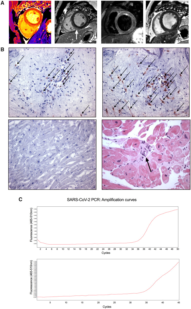
Biopsy-proven SARS-CoV-2 mRNA in clinically suspected myocarditis. (A) Representative cardiac magnetic resonance imaging (cMRI) scans. The two left panels: patient A—native T1 map showing prolonged T1 relaxation times in the posterior interventricular septum and corresponding late gadolinium enhancement image (LGE) with enhancement in the posterior septum (arrowhead), consistent with acute myocarditis. Transthoracic echocardiography showed a preserved left ventricular (LV) systolic function (EF 60%) without any wall motion abnormalities, but focal echo-bright appearance of the interventricular septum (not shown) and slightly impaired global longitudinal strain. The two right panels: patient B—representative cMRI scans of the patient who had a history of coronary artery disease treated by percutaneous coronary intervention (everolimus-eluting stent in the right coronary artery). T2-short TI inversion recovery image, showing diffuse myocardial oedema and LGE image with subtle subepicardial enhancement of the lateral wall (arrowheads). Transthoracic echocardiography showed LV dysfunction (EF 30%), decreased global and regional longitudinal strain, as well as increased LV end-diastolic diameter. (B) Representative immunohistochemical staining for assessment of inflammation in SARS-CoV-2-positive EMB (patient B). Top left panel: increased CD3+ T lymphocytes. Infiltrates of inflammatory cells (arrowheads) mostly in the neighbourhood of small blood vessels. Top right panel: increased CD45R0+ T memory (arrowheads) cells mostly in the neighbourhood of small blood vessels. Bottom left panel: negative control of CD3 immunostaining. Magnification ×200. Bottom right panel: histological evaluations were performed on paraffin sections with haematoxylin and eosin (HE; patient A). The arrow indicates increased thickness of the small arterial vessel. No active myocarditis according to Dallas criteria (‘borderline myocarditis’). Magnification ×400. (C) Expression analysis of SARS-CoV-2-specific nucleic acid was performed by an RT–PCR assay (TIB MOLBIOL, Roche, Germany) in cardiac tissue obtained by EMB. Original amplification curves of patient A (top) and patient B (bottom). See also Supplementary material online Table S1 and Figure S1.
References
-
- Huang C, Wang Y, Li X, Ren L, Zhao J, Hu Y, Zhang L, Fan G, Xu J, Gu X, Cheng Z, Yu T, Xia J, Wei Y, Wu W, Xie X, Yin W, Li H, Liu M, Xiao Y, Gao H, Guo L, Xie J, Wang G, Jiang R, Gao Z, Jin Q, Wang J, Cao B.. Clinical features of patients infected with 2019 novel coronavirus in Wuhan, China. Lancet 2020;395:497–506. - PMC - PubMed
-
- Guzik TJ, Mohiddin SA, Dimarco A, Patel V, Savvatis K, Marelli-Berg FM, Madhur MS, Tomaszewski M, Maffia P, D’Acquisto F, Nicklin SA, Marian AJ, Nosalski R, Murray EC, Guzik B, Berry C, Touyz RM, Kreutz R, Wang DW, Bhella D, Sagliocco O, Crea F, Thomson EC, McInnes IB.. COVID-19 and the cardiovascular system: implications for risk assessment, diagnosis, and treatment options. Cardiovasc Res 2020;doi:10.1093/cvr/cvaa106 - PMC - PubMed
-
- Tavazzi G, Pellegrini C, Maurelli M, Belliato M, Sciutti F, Bottazzi A, Sepe PA, Resasco T, Camporotondo R, Bruno R, Baldanti F, Paolucci S, Pelenghi S, Iotti GA, Mojoli F, Arbustini E.. Myocardial localization of coronavirus in COVID-19 cardiogenic shock. Eur J Heart Fail 2020;22:911–915. - PMC - PubMed
Publication types
MeSH terms
Substances
LinkOut - more resources
Full Text Sources
Miscellaneous
