

SARS-CoV-2 endothelial infection causes COVID-19 chilblains: histopathological, immunohistochemical and ultrastructural study of seven paediatric cases
- PMID: 32562567
- PMCID: PMC7323219
- DOI: 10.1111/bjd.19327
SARS-CoV-2 endothelial infection causes COVID-19 chilblains: histopathological, immunohistochemical and ultrastructural study of seven paediatric cases
Abstract
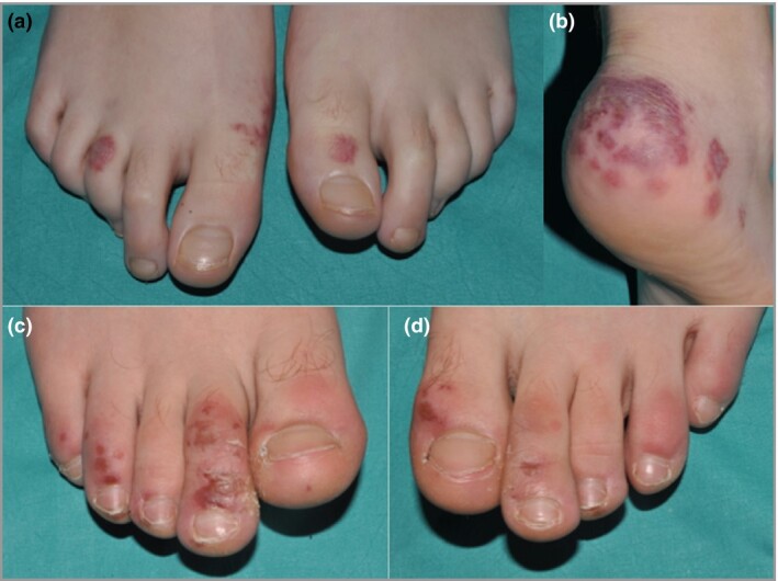
Background: Chilblains ('COVID toes') are being seen with increasing frequency in children and young adults during the COVID-19 pandemic. Detailed histopathological descriptions of COVID-19 chilblains have not been reported, and causality of SARS-CoV-2 has not yet been established.
Objectives: To describe the histopathological features of COVID-19 chilblains and to explore the presence of SARS-CoV-2 in the tissue.
Methods: We examined skin biopsies from seven paediatric patients presenting with chilblains during the COVID-19 pandemic. Immunohistochemistry for SARS-CoV-2 was performed in all cases and electron microscopy in one.
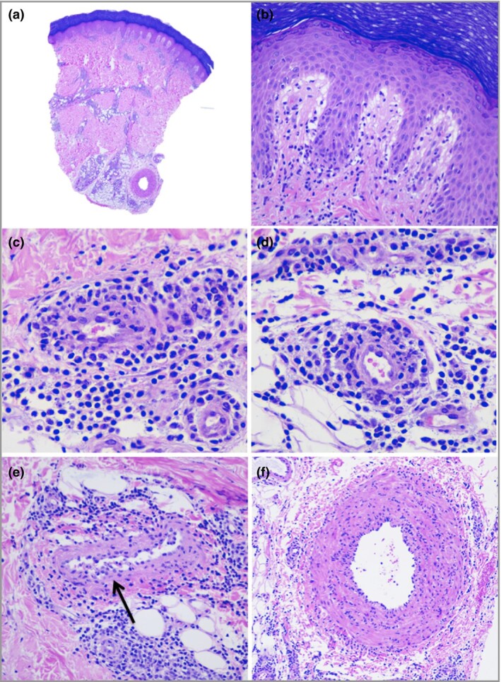
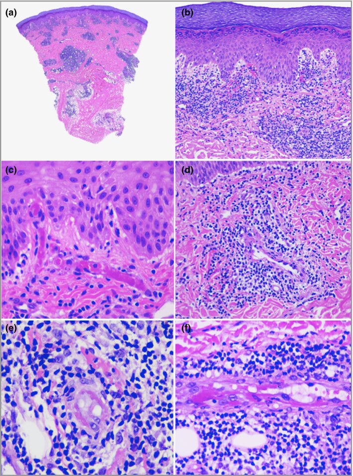
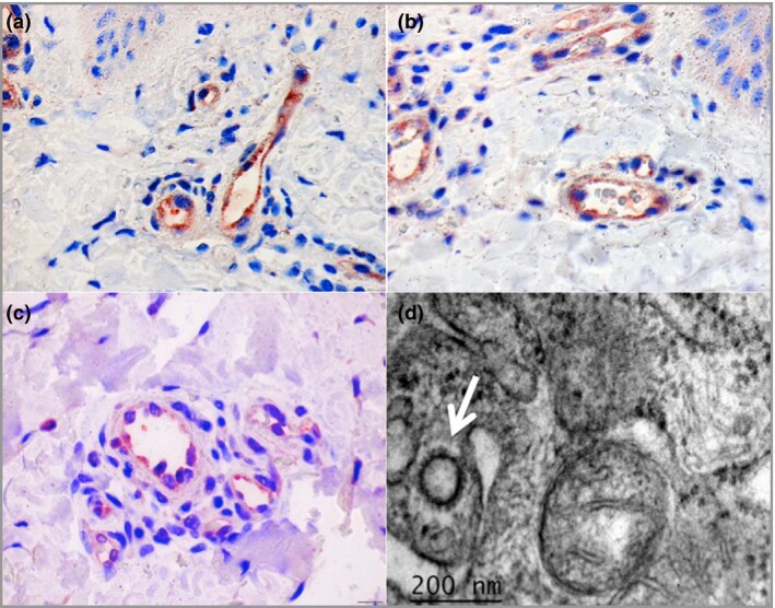
Results: Histopathology showed variable degrees of lymphocytic vasculitis ranging from endothelial swelling and endotheliitis to fibrinoid necrosis and thrombosis. Purpura, superficial and deep perivascular lymphocytic inflammation with perieccrine accentuation, oedema, and mild vacuolar interface damage were also seen. SARS-CoV-2 immunohistochemistry was positive in endothelial cells and epithelial cells of eccrine glands. Coronavirus particles were found in the cytoplasm of endothelial cells on electron microscopy.
Conclusions: Although the clinical and histopathological features were similar to other forms of chilblains, the presence of viral particles in the endothelium and the histological evidence of vascular damage support a causal relation of the lesions with SARS-CoV-2. Endothelial damage induced by the virus could be the key mechanism in the pathogenesis of COVID-19 chilblains and perhaps also in a group of patients severely affected by COVID-19 presenting with features of microangiopathic damage. What is already known about this topic? Despite the high number of cases of chilblains seen during the COVID-19 pandemic, a definite causative role for SARS-CoV-2 has not yet been proven. Different pathogenetic hypotheses have been proposed, including coagulation anomalies, interferon release and external factors. What does this study add? The demonstration of SARS-CoV-2 in endothelial cells of skin biopsies by immunohistochemistry and electron microscopy confirms that these lesions are part of the spectrum of COVID-19. Virus-induced vascular damage and secondary ischaemia could explain the pathophysiology of COVID-19 chilblains. Our findings support the hypothesis that widespread endothelial infection by SARS-CoV-2 could have a pathogenetic role in the severe forms of COVID-19. Linked Comment: Wetter. Br J Dermatol 2020; 183:611.
© 2020 British Association of Dermatologists.
Figures
Comment in
-
Chilblains and COVID-19: why SARS-CoV-2 endothelial infection is questioned.Br J Dermatol. 2020 Dec;183(6):1152-1153. doi: 10.1111/bjd.19489. Epub 2020 Sep 24. Br J Dermatol. 2020. PMID: 32798309 Free PMC article.
-
SARS-CoV-2 PCR testing of skin for COVID-19 diagnostics: a case report.Lancet. 2020 Aug 29;396(10251):598-599. doi: 10.1016/S0140-6736(20)31754-2. Epub 2020 Aug 13. Lancet. 2020. PMID: 32798450 Free PMC article. No abstract available.
-
Illuminating, through immunohistochemistry, the link between SARS-CoV-2 and pernio (chilblains).Br J Dermatol. 2020 Oct;183(4):611. doi: 10.1111/bjd.19405. Epub 2020 Sep 1. Br J Dermatol. 2020. PMID: 32869311 Free PMC article.
-
SARS-CoV-2 has not been detected directly by electron microscopy in the endothelium of chilblain lesions.Br J Dermatol. 2021 Jan;184(1):186. doi: 10.1111/bjd.19572. Epub 2020 Dec 16. Br J Dermatol. 2021. PMID: 33000462 Free PMC article.
References
-
- BBC News. Coronavirus: ‘Covid toe’ and other rashes puzzle doctors. Available at: https://www.bbc.com/news/health‐52493574 (last accessed 20 July 2020).
MeSH terms
LinkOut - more resources
Full Text Sources
Medical
Research Materials
Miscellaneous
