Review
. 2020 Sep:163:103624.
doi: 10.1016/j.mod.2020.103624. Epub 2020 Jun 17.

Cellular processes driving gastrulation in the avian embryo

Affiliations
Review

Cellular processes driving gastrulation in the avian embryo

Guillermo Serrano Nájera et al. Mech Dev. 2020 Sep.

Abstract

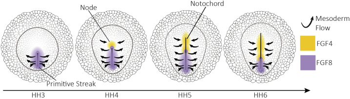
Gastrulation consists in the dramatic reorganisation of the epiblast, a one-cell thick epithelial sheet, into a multilayered embryo. In chick, the formation of the internal layers requires the generation of a macroscopic convection-like flow, which involves up to 50,000 epithelial cells in the epiblast. These cell movements locate the mesendoderm precursors into the midline of the epiblast to form the primitive streak. There they acquire a mesenchymal phenotype, ingress into the embryo and migrate outward to populate the inner embryonic layers. This review covers what is currently understood about how cell behaviours ultimately cause these morphogenetic events and how they are regulated. We discuss 1) how the biochemical patterning of the embryo before gastrulation creates compartments of differential cell behaviours, 2) how the global epithelial flows arise from the coordinated actions of individual cells, 3) how the cells delaminate individually from the epiblast during the ingression, and 4) how cells move after the ingression following stereotypical migration routes. We conclude by exploring new technical advances that will facilitate future research in the chick model system.

Keywords: Cell flows; Chick embryo; Gastrulation; Intercalation; Morphogenesis; Patterning.


Conflict of interest statement

Declaration of competing interest The authors have no competing interests to declare.

Figures

Fig. 1
Fig. 1
Avian development before gastrulation. A) Orientation of the embryo on the yolk during intrauterine stages of development. Arrows indicate the direction of rotation of the egg and yolk during the transition through the oviduct. B) Segregation of the embryonic epiblast and extra embryonic Area Opaca. C) Development of the hypoblast. D) Cross sections during intrauterine development. E) Spatial patterning of essential paracrine developmental signals: Wnt8A is initially expressed in the Area Opaca, there is an anterior-posterior gradient of Bmp4 in the Area Opaca, Gdf3 is expressed in the posterior epiblast and Fgf8 is produced in the hypoblast.
Fig. 2
Fig. 2
Tissue flows driving streak formation. A) Tissue flow pattern in the embryo (white arrows). Every cell represents ~50 cells in the real embryo. B) Isotropic and anisotropic components of the strain rates derived from the tissue flow. The isotropic component results from cell growth and ingression while the anisotropic component is the product of asymmetric cell rearrangements and cell shape changes. C) Cross sections during streak formation. Cells in the mesendoderm become increasingly columnar as they approach the midline. After digestion of the basement membrane, mesendoderm precursor cells undergo EMT and migrate out to populate the inner embryonic layers.
Fig. 3
Fig. 3
Origins of flow organisation. A) Flow organisers. From left to right: Attractors and repellers of the tissue flow, territories of directed and stochastic cell intercalation and myosin cables B) Cell behaviours that drive epithelial tissue flows, cell intercalation, cell division and ingressions. Division and ingression also contribute to cell rearrangements since they create changes of epithelial topology. C) Mechanism of contraction propagation based on tension dependent stabilisation of apical junctional myosin. From top to bottom: 1) The apical contraction of an individual cell due to a local accumulation of myosin, 2) results in the stretching of neighbouring cell junctions. The increase in tension produced by the stretching leads to the local stabilisation of myosin in these cells resulting in 3) apical contraction in these cells and propagation to further neighbours.
Fig. 4
Fig. 4
Mechanism of cell ingression. Before ingression, cells need to contract apically and disassemble tight and adherens junctions with their neighbours. During the process of ingression, cells get traction from the neighbour cells, extend protrusions and slide down, presumably, using a retrograde actin flow (black arrows). Finally, the cell acquires a mesenchymal phenotype and has converted the apicobasal polarity into a front-back polarity.
Fig. 5
Fig. 5
FGF signalling in mesoderm migration. 1) FGF8 is secreted in the primitive streak and act as a repulsive signal for the mesoderm cells. Once cells ingress they migrate out via chemo-repulsion. 2) When the node starts to regress it creates the notochord which secretes FGF4 and attracts migrant mesoderm cells back towards the midline.

Similar articles

Cited by

References

    1. Acloque H., Ocana O.H., Matheu A., Rizzoti K., Wise C., Lovell-Badge R., Nieto M.A. Reciprocal repression between Sox3 and Snail transcription factors defines embryonic territories at gastrulation. Dev. Cell. 2011;21:546–558. - PMC - PubMed
    1. Acloque H., Ocana O.H., Abad D., Stern C.D., Nieto M.A. Snail2 and Zeb2 repress P-cadherin to define embryonic territories in the chick embryo. Development. 2017;144:649–656. - PubMed
    1. Alev C., Wu Y.P., Nakaya Y., Sheng G.J. Decoupling of amniote gastrulation and streak formation reveals a morphogenetic unity in vertebrate mesoderm induction. Development. 2013;140:2691–2696. - PubMed
    1. Arendt D., Nubler-Jung K. Rearranging gastrulation in the name of yolk: evolution of gastrulation in yolk-rich amniote eggs. Mech. Dev. 1999;81:3–22. - PubMed
    1. Arias C.F., Herrero M.A., Stern C.D., Bertocchini F. A molecular mechanism of symmetry breaking in the early chick embryo. Sci. Rep. 2017;7 - PMC - PubMed

Publication types

LinkOut - more resources