

Mammographic density changes during neoadjuvant breast cancer treatment: NeoDense, a prospective study in Sweden
- PMID: 32563178
- PMCID: PMC7375568
- DOI: 10.1016/j.breast.2020.05.013
Mammographic density changes during neoadjuvant breast cancer treatment: NeoDense, a prospective study in Sweden
Abstract
Objectives: To assess if mammographic density (MD) changes during neoadjuvant breast cancer treatment and is predictive of a pathological complete response (pCR).
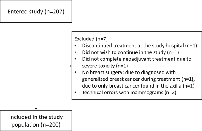
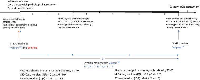
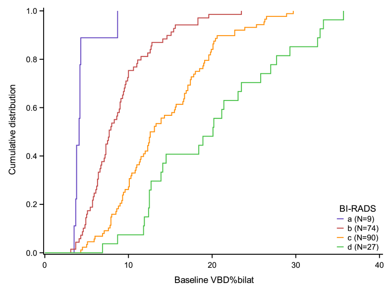
Methods: We prospectively included 200 breast cancer patients assigned to neoadjuvant chemotherapy (NACT) in the NeoDense study (2014-2019). Raw data mammograms were used to assess MD with a fully automated volumetric method and radiologists categorized MD using the Breast Imaging-Reporting and Data System (BI-RADS), 5th Edition. Logistic regression was used to calculate odds ratios (OR) for pCR comparing BI-RADS categories c vs. a, b, and d as well as with a 0.5% change in percent dense volume adjusting for baseline characteristics.
Results: The overall median age was 53.1 years, and 48% of study participants were premenopausal pre-NACT. A total of 23% (N = 45) of the patients accomplished pCR following NACT. Patients with very dense breasts (BI-RADS d) were more likely to have a positive axillary lymph node status at diagnosis: 89% of the patients with very dense breasts compared to 72% in the entire cohort. A total of 74% of patients decreased their absolute dense volume during NACT. The likelihood of accomplishing pCR following NACT was independent of volumetric MD at diagnosis and change in volumetric MD during treatment. No trend was observed between decreasing density according to BI-RADS and the likelihood of accomplishing pCR following NACT.
Conclusions: The majority of patients decreased their MD during NACT. We found no evidence of MD as a predictive marker of pCR in the neoadjuvant setting.
Keywords: Breast cancer; Breast density; Mammography; Neoadjuvant therapy.
Copyright © 2020 The Author(s). Published by Elsevier Ltd.. All rights reserved.
Conflict of interest statement
Declaration of competing interest SZ and HS have received speakers’ fees and travel support from Siemens Healthcare AG. SZ has received consultancy fees from Collective Minds Radiology AB. PH is a member of a scientific advisory board for: Cancer Research UK, iCAD and Atossa Genetics. SB has received speakers’ fees from Pfizer, is a member of a Pfizer advisory board, and has received travel support from Roche. The other authors declare that they have no competing interests.
Figures
References
-
- McCormack V.A., dos Santos Silva I. Breast density and parenchymal patterns as markers of breast cancer risk: a meta-analysis. Cancer Epidemiol Biomark Prev. 2006;15(6):1159–1169. - PubMed
-
- Sickles E., D’Orsi C.J., Bassett L.W. ACR BI-RADS® Atlas, breast imaging reporting and data system. American College of Radiology; Reston, VA: 2013. ACR BI-RADS® mammography.
MeSH terms
Substances
LinkOut - more resources
Full Text Sources
Medical
