. 2020 Aug 4;222(5):734-745.
doi: 10.1093/infdis/jiaa356.

Attenuated Interferon and Proinflammatory Response in SARS-CoV-2-Infected Human Dendritic Cells Is Associated With Viral Antagonism of STAT1 Phosphorylation

Affiliations

Attenuated Interferon and Proinflammatory Response in SARS-CoV-2-Infected Human Dendritic Cells Is Associated With Viral Antagonism of STAT1 Phosphorylation

Dong Yang et al. J Infect Dis. .

Abstract

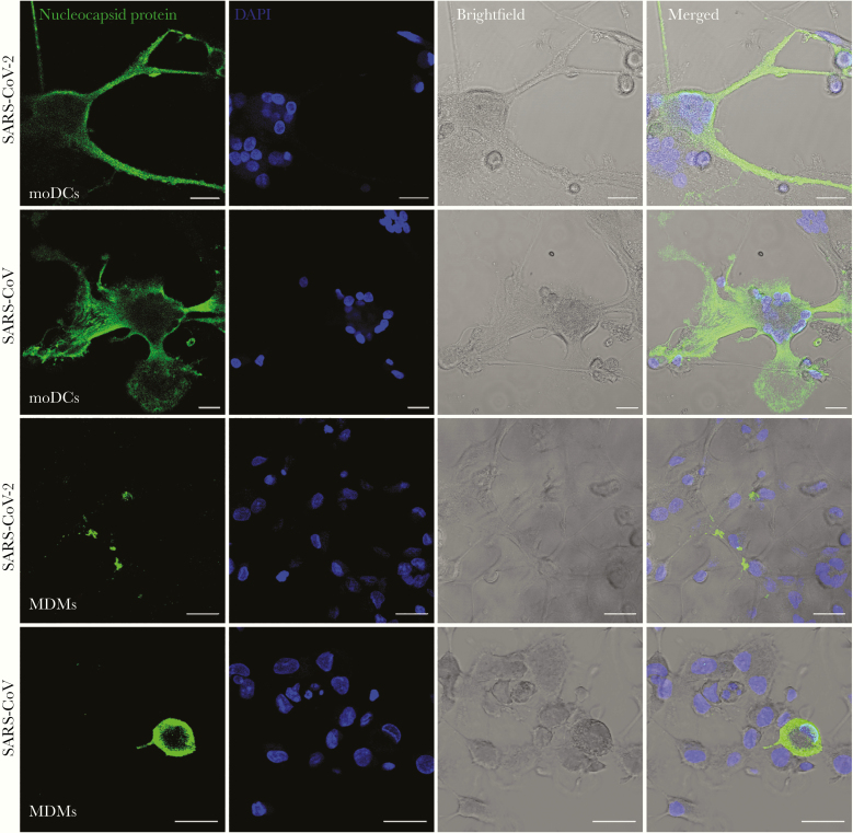
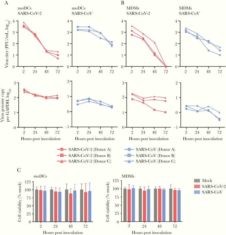
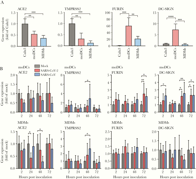
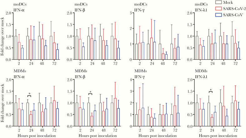
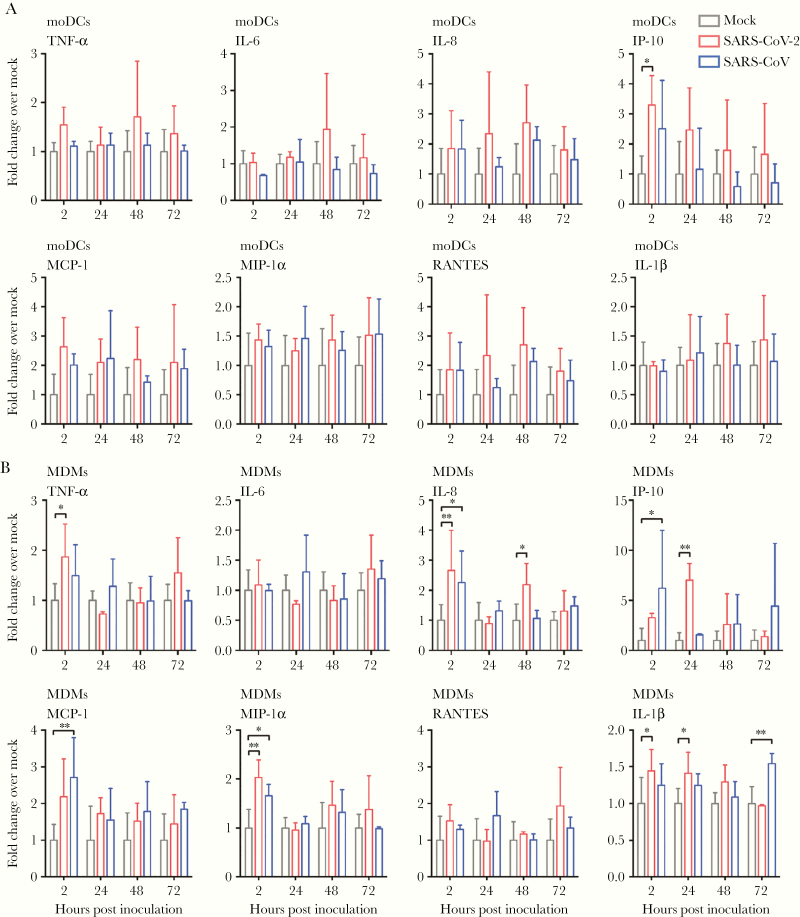
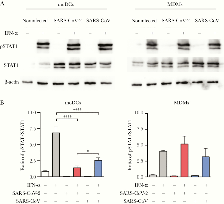
Clinical manifestations of coronavirus disease 2019 (COVID-19) vary from asymptomatic virus shedding, nonspecific pharyngitis, to pneumonia with silent hypoxia and respiratory failure. Dendritic cells and macrophages are sentinel cells for innate and adaptive immunity that affect the pathogenesis of severe acute respiratory syndrome (SARS) and Middle East respiratory syndrome (MERS). The interplay between SARS-CoV-2 and these cell types remains unknown. We investigated infection and host responses of monocyte-derived dendritic cells (moDCs) and macrophages (MDMs) infected by SARS-CoV-2. MoDCs and MDMs were permissive to SARS-CoV-2 infection and protein expression but did not support productive virus replication. Importantly, SARS-CoV-2 launched an attenuated interferon response in both cell types and triggered significant proinflammatory cytokine/chemokine expression in MDMs but not moDCs. Investigations suggested that this attenuated immune response to SARS-CoV-2 in moDCs was associated with viral antagonism of STAT1 phosphorylation. These findings may explain the mild and insidious course of COVID-19 until late deterioration.

Keywords: COVID-19; MDMs; SARS-CoV-2; coronavirus; dendritic cells; macrophages; moDCs.


Figures

Figure 1.
Figure 1.
MoDCs and MDMs were susceptible to SARS-CoV-2 infection. MoDCs and MDMs were challenged with SARS-CoV-2 or SARS-CoV at a MOI of 10. At 24 hours post inoculation, the cells were fixed in 4% paraformaldehyde, permeabilized, and incubated with an in-house rabbit antiserum against SARS-CoV-2 nucleocapsid protein, followed by incubation with Alexa Fluor 488 secondary antibody for 1 hour. The slides were imaged with confocal microscopy using a Carl Zeiss LSM 880 system. Bars represent 20 μm. Abbreviations: MOI, multiplicity of infection; DAPI, 4′,6-diamidino-2-phenylindole; moDCs, monocyte-derived dendritic cells; MDMs, monocyte-derived macrophages; SARS-CoV-2, severe acute respiratory syndrome coronavirus 2.
Figure 2.
Figure 2.
Infection of SARS-CoV-2 in moDCs and MDMs was not productive. MoDCs (A) and MDMs (B) were infected with SARS-CoV-2 or SARS-CoV at an MOI of 1. The live infectious virus particles in the supernatants and the viral genome copy in the cell lysates were determined by plaque assays and qRT-PCR, respectively. C, The cell viability of moDCs and MDMs upon SARS-CoV-2 or SARS-CoV infection at an MOI of 1 was quantified at the indicated hours post infection using CellTiterGlo assays. The mean cell viability of SARS-CoV-2– or SARS-CoV–infected cells was compared with that of mock-infected cells at each time point. The results represent mean and standard deviations from 3 individual donors in 3 independent experiments. Statistical significance between the groups was determined with 1-way ANOVA and was consider significant when P < .05. Abbreviations: MOI, multiplicity of infection; PFU, plaque-forming unit; GAPDH, glyceraldehyde-3-phosphate dehydrogenase; moDCs, monocyte-derived dendritic cells; MDMs, monocyte-derived macrophages; qRT-PCR, quantitative reverse transcription polymerase chain reaction; SARS-CoV-2, severe acute respiratory syndrome coronavirus 2.
Figure 3.
Figure 3.
SARS-CoV-2 modulated the differential expression of entry-related host factors in moDCs and MDMs. A, The expression of endogenous ACE2, TMPRSS2, FURIN, and DC-SIGN was evaluated in moDCs and MDMs. Calu3 cells were included as a positive control. B, MoDCs and MDMs were infected with SARS-CoV-2 or SARS-CoV at an MOI of 1. The cell lysates were harvested to detect expression of entry-related host factors using qRT-PCR at the indicated hours post infection. The expression of entry-related host factors in SARS-CoV-2–infected or SARS-CoV–infected cells was compared with that of mock-infected cells at each time point. The results represent mean and standard deviations from 3 to 6 individual donors in 3 independent experiments. Statistical significance between groups was determined with 1-way ANOVA and was consider significant when P < .05. * P < .05, ** P < .01, *** P < .001, **** P < .0001. Abbreviations: MOI, multiplicity of infection; ACE2, angiotensin-converting enzyme 2; TMPRSS2; transmembrane protease serine 2; DC-SIGN, dendritic cell-specific intercellular adhesion molecule-3-grabbing non-integrin; moDCs, monocyte-derived dendritic cells; MDMs, monocyte-derived macrophages; qRT-PCR, quantitative reverse transcription polymerase chain reaction; SARS-CoV-2, severe acute respiratory syndrome coronavirus 2.
Figure 4.
Figure 4.
Attenuated IFN response in SARS-CoV-2–infected moDCs and MDMs. MoDCs and MDMs were inoculated with SARS-CoV-2 or SARS-CoV at an MOI of 1. The cell lysates were harvested for qRT-PCR analysis of type I (IFN-α and IFN-β), II (IFN-γ), and III (IFN-λ1) IFN. The results represent mean and standard deviations from 3 to 6 individual donors in 3 independent experiments. Statistical significance between the groups was determined with 2-way ANOVA and was consider significant when P < .05. * P < .05. Abbreviations: MOI, multiplicity of infection; IFN, interferon; moDCs, monocyte-derived dendritic cells; MDMs, monocyte-derived macrophages; qRT-PCR, quantitative reverse transcription polymerase chain reaction; SARS-CoV-2, severe acute respiratory syndrome coronavirus 2.
Figure 5.
Figure 5.
SARS-CoV-2 induced significant proinflammatory response in MDMs but not moDCs. MoDCs (A) and MDMs (B) were inoculated with SARS-CoV-2 or SARS-CoV at an MOI of 1. The cell lysates were harvested for qRT-PCR analysis of representative proinflammatory cytokines and chemokines. The results represent mean and standard deviations from 3 to 6 individual donors in 3 independent experiments. Statistical significance between the groups was determined with 2-way ANOVA and was consider significant when P < .05. * P < .05, ** P < .01. Abbreviations: MOI, multiplicity of infection; TNF-α, tumor necrosis factor-α; IP-10, IFN-γ inducible protein-10; MCP-1, monocyte chemoattractant protein-1; MIP-1α, macrophage-inflammatory protein-1α; RANTES, regulated upon activation normal T-cell expressed and secreted; moDCs, monocyte-derived dendritic cells; MDMs, monocyte-derived macrophages; qRT-PCR, quantitative reverse transcription polymerase chain reaction; SARS-CoV-2, severe acute respiratory syndrome coronavirus 2.
Figure 6.
Figure 6.
SARS-CoV-2 antagonized STAT1 phosphorylation in moDCs. MoDCs and MDMs were mock-infected or infected with SARS-CoV-2 or SARS-CoV at an MOI of 10. At 24 hours post infection, the cells were untreated or treated with 1000 U/mL of recombinant human IFN-α for 40 minutes. The cell lysates were collected for the detection of STAT1, pSTAT1, and β-actin by Western blots. A, Representative blots are shown from 3 donors in 3 independent experiments. B, Quantitation was calculated as the ratio of pSTAT1 over STAT1 protein. Statistical analysis was performed with 1-way ANOVA and the differences were considered significant when P < .05. *P < .05, ****P < .0001. Abbreviations: MOI, multiplicity of infection; IFN, interferon; moDCs, monocyte-derived dendritic cells; MDMs, monocyte-derived macrophages; SARS-CoV-2, severe acute respiratory syndrome coronavirus 2.

References

    1. Zhu N, Zhang D, Wang W, et al. . A novel coronavirus from patients with pneumonia in China, 2019. N Engl J Med 2020; 382:727–33. - PMC - PubMed
    1. Chan JF, Yuan S, Kok KH, et al. . A familial cluster of pneumonia associated with the 2019 novel coronavirus indicating person-to-person transmission: a study of a family cluster. Lancet 2020; 395:514–23. - PMC - PubMed
    1. Chan JF, Kok KH, Zhu Z, et al. . Genomic characterization of the 2019 novel human-pathogenic coronavirus isolated from a patient with atypical pneumonia after visiting Wuhan. Emerg Microbes Infect 2020; 9:221–36. - PMC - PubMed
    1. World Health Organization. Coronavirus disease (COVID-19) situation report–108 https://www.who.int/docs/default-source/coronaviruse/situation-reports/2.... Accessed 23 June 2020.
    1. Guan WJ, Ni ZY, Hu Y, et al. . Clinical characteristics of coronavirus disease 2019 in China. N Engl J Med 2020; 382:1708–20. - PMC - PubMed

Publication types

MeSH terms