Review
. 2020 Jun 20;19(1):107.
doi: 10.1186/s12943-020-01227-0.

PARP inhibitor resistance: the underlying mechanisms and clinical implications

Affiliations
Review

PARP inhibitor resistance: the underlying mechanisms and clinical implications

He Li et al. Mol Cancer. .

Abstract

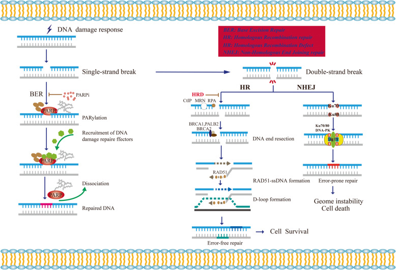
Due to the DNA repair defect, BRCA1/2 deficient tumor cells are more sensitive to PARP inhibitors (PARPi) through the mechanism of synthetic lethality. At present, several PAPRi targeting poly (ADP-ribose) polymerase (PARP) have been approved for ovarian cancer and breast cancer indications. However, PARPi resistance is ubiquitous in clinic. More than 40% BRCA1/2-deficient patients fail to respond to PARPi. In addition, lots of patients acquire PARPi resistance with prolonged oral administration of PARPi. Homologous recombination repair deficient (HRD), as an essential prerequisite of synthetic lethality, plays a vital role in killing tumor cells. Therefore, Homologous recombination repair restoration (HRR) becomes the predominant reason of PARPi resistance. Recently, it was reported that DNA replication fork protection also contributed to PARPi resistance in BRCA1/2-deficient cells and patients. Moreover, various factors, such as reversion mutations, epigenetic modification, restoration of ADP-ribosylation (PARylation) and pharmacological alteration lead to PARPi resistance as well. In this review, we reviewed the underlying mechanisms of PARP inhibitor resistance in detail and summarized the potential strategies to overcome PARPi resistance and increase PARPi sensitivity.

Keywords: Homologous recombination; PARPi; Resistance; Synthetic lethality.


Conflict of interest statement

The authors declare that there is no potential competing interest.

Figures

Fig. 1
Fig. 1
Schematic describing the function the principle of synthetic lethality interaction between PARPs and BRCA1/2. When cells suffer from DNA response, single-strand breaks emerge. PARPs, especially PARP1, bind to the DNA break sites, which result in the PARylation of target proteins and recrement of the DNA damage repair effectors. Then the auto-PARylation on PARPs leads to the dissociation of PARPs from DNA. Treating HR-deficient tumor cells with PARPi, NHEJ is the only pathway to use to repair double-strand break, which lead to accumulation of genome instability and cell death for the low fidelity
Fig. 2
Fig. 2
Homologous recombination repair in S/G2 phase. The double-strand break ends are resected by MRE11-RAD50-NBS1(MRN) complex together with CtIP. ATM is recruited to DSBs through MRN and phosphorylates targets such as 53BP1 and MDC1. MDC1 phosphorylation recruits the E3 ubiquitin ligase RNF8, which, through recruitment of a second E3 ubiquitin ligase (RNF168), leads to histone H2A ubiquitylation. This modification, together with H4K20 methylation, allows for 53BP1 recruitment. 53BP1 phosphorylation allows its interaction with RIF1 and PTIP, which can be blocked by WIP1. 53BP1 blocks DNA resection by recruiting shieldin and presents cells to NHEJ. While, BRCA1 counteracts the protection function of 53BP1, leading to the resection of DNA ends. Afterwards, the resected DNA ends are coated by PRA. With the favor of PALB2, BRCA2 binds with BRCA1 and promotes the loading of RAD51. The RAD51 mediates the invasion of the homologous sequence and formation of the nucleoprotein filament and D-loop by eliminating secondary structure formation. EMI and DDB2 mediate the degradation of RAD51. TOPBP1 phosphorylates RAD51. BRD4 and HORMAD1 are key regulators of RAD51 accumulation on chromatin. P, phosphorylation; Ub, ubiquitylation; Me, methylation, SUMO, SUMOylation, red arrows, resection
Fig. 3
Fig. 3
Schematic describing the function of PARP1 and BRCA1/2 in protection of DNA replication fork and mechanisms of protection of DNA replication fork leading to PARPi resistance. a PARPi trap PARP1 on DNA and cause fork stalling. After fork stalling, PRA is phosphorylated and ssDNA is coated by PRA rapidly. Then, RAD51 replaces RPA and mediates replication fork reversal. The revered fork can be degraded by MRE11 and MUS81. BRCA1/2 is relied on to protect nascent DNA replication forks from degradation. EZH2 induces H3K27 methylation and MUS81 recruitment at stalled forks. MELL3/4 induces H3K4 methylation increases the accumulation of PTIP, which leads to the recruitment of MRE11. FANCD2 suppresses MRE11-mediated fork degradation, which can be reversed by SMARCAL1. RADX blocks the recruitment of RAD51 at replication forks. b In BRCA1/2-deficient cells, low expression of PARP1, RADX, SMARCAL1, EZH2, PTIP, MELL3/4 and high expression of FANCD2 confer resistance of PARPi

References

    1. Jeggo PA, Pearl LH, Carr AM. DNA repair, genome stability and cancer: a historical perspective. Nat Rev Cancer. 2016;16:35–42. - PubMed
    1. Chatterjee N, Walker GC. Mechanisms of DNA damage, repair, and mutagenesis. Environ Mol Mutagen. 2017;58:235–263. - PMC - PubMed
    1. Langelier M-F, Riccio AA, Pascal JM. PARP-2 and PARP-3 are selectively activated by 5′ phosphorylated DNA breaks through an allosteric regulatory mechanism shared with PARP-1. Nucleic Acids Res. 2014;42:7762–7775. - PMC - PubMed
    1. Eustermann S, Wu W-F, Langelier M-F, Yang J-C, Easton LE, Riccio AA, Pascal JM, Neuhaus D. Structural basis of detection and signaling of DNA single-Strand breaks by human PARP-1. Mol Cell. 2015;60:742–754. - PMC - PubMed
    1. Langelier M-F, Planck JL, Roy S, Pascal JM. Structural basis for DNA damage-dependent poly(ADP-ribosyl) ation by human PARP-1. Science (New York, NY) 2012;336:728–732. - PMC - PubMed

Publication types

Substances