

Pathological Findings in the Testes of COVID-19 Patients: Clinical Implications
- PMID: 32563676
- PMCID: PMC7261470
- DOI: 10.1016/j.euf.2020.05.009
Pathological Findings in the Testes of COVID-19 Patients: Clinical Implications
Abstract
Background: Coronavirus disease 2019 (COVID-19), caused by severe acute respiratory syndrome coronavirus 2 (SARS-CoV-2), involves multiple organs. Testicular involvement is largely unknown.
Objective: To determine the pathological changes and whether SARS-CoV-2 can be detected in the testes of deceased COVID-19 patients.
Design, setting, and participants: Postmortem examination of the testes from 12 COVID-19 patients was performed using light and electron microscopy, and immunohistochemistry for lymphocytic and histiocytic markers. Reverse transcription-polymerase chain reaction (RT-PCR) was used to detect the virus in testicular tissue.
Outcome measurements and statistical analysis: Seminiferous tubular injury was assessed as none, mild, moderate, or severe according to the extent of tubular damage. Leydig cells in the interstitium were counted in ten 400× microscopy fields.
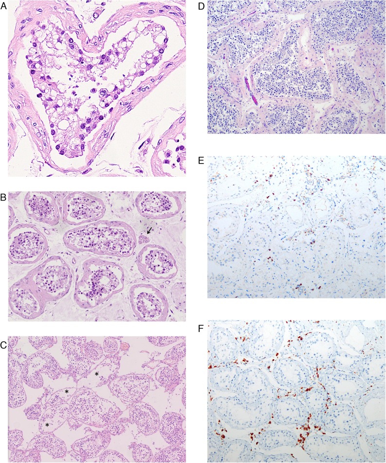
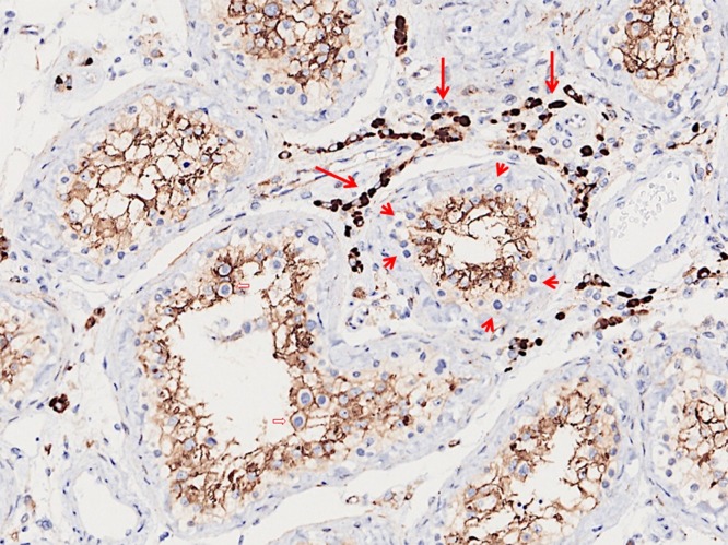
Results and limitations: Microscopically, Sertoli cells showed swelling, vacuolation and cytoplasmic rarefaction, detachment from tubular basement membranes, and loss and sloughing into lumens of the intratubular cell mass. Two, five, and four of 11 cases showed mild, moderate, and severe injury, respectively. The mean number of Leydig cells in COVID-19 testes was significantly lower than in the control group (2.2 vs 7.8, p < 0.001). In the interstitium there was edema and mild inflammatory infiltrates composed of T lymphocytes and histiocytes. Transmission EM did not identify viral particles in three cases. RT-PCR detected the virus in one of 12 cases.
Conclusions: Testes from COVID-19 patients exhibited significant seminiferous tubular injury, reduced Leydig cells, and mild lymphocytic inflammation. We found no evidence of SARS-CoV-2 virus in the testes in the majority (90%) of the cases by RT-PCR, and in none by electron microscopy. These findings can provide evidence-based guidance for sperm donation and inform management strategies to mitigate the risk of testicular injury during the COVID-19 disease course.
Patient summary: We examined the testes of deceased COVID-19 patients. We found significant damage to the testicular parenchyma. However, virus was not detected in testes in the majority of cases.
Keywords: COVID-19; Fertility; Postmortem needle autopsy; SARS-CoV-2; Testis.
Copyright © 2020 European Association of Urology. Published by Elsevier B.V. All rights reserved.
Figures
References
-
- World Health Organization. Coronavirus disease 2019 (COVID-19) situation report 105. www.who.int/docs/default-source/coronaviruse/situation-reports/20200504-....
-
- Yao X.H., Li T.Y., He Z.C. A pathological report of three COVID-19 cases by minimally invasive autopsies. Zhonghua Bing Li Xue Za Zhi. 2020;49:E009. - PubMed
MeSH terms
Substances
LinkOut - more resources
Full Text Sources
Medical
Miscellaneous
