Review
. 2020 Oct;38(10):1066-1098.
doi: 10.1016/j.tibtech.2020.05.012. Epub 2020 Jun 18.

Technologies and Standardization in Research on Extracellular Vesicles

Affiliations
Review

Technologies and Standardization in Research on Extracellular Vesicles

Srujan Gandham et al. Trends Biotechnol. 2020 Oct.

Abstract

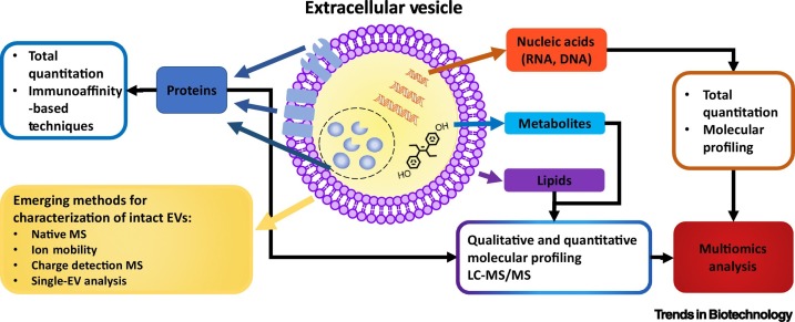
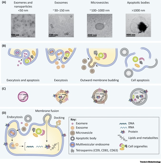
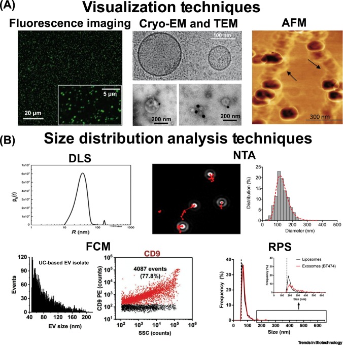
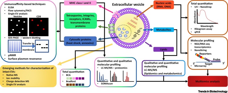
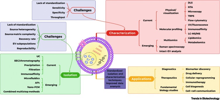
Extracellular vesicles (EVs) are phospholipid bilayer membrane-enclosed structures containing RNAs, proteins, lipids, metabolites, and other molecules, secreted by various cells into physiological fluids. EV-mediated transfer of biomolecules is a critical component of a variety of physiological and pathological processes. Potential applications of EVs in novel diagnostic and therapeutic strategies have brought increasing attention. However, EV research remains highly challenging due to the inherently complex biogenesis of EVs and their vast heterogeneity in size, composition, and origin. There is a need for the establishment of standardized methods that address EV heterogeneity and sources of pre-analytical and analytical variability in EV studies. Here, we review technologies developed for EV isolation and characterization and discuss paths toward standardization in EV research.

Keywords: characterization; exosomes; extracellular vesicles; isolation; molecular profiling; standardization.


Figures

Figure 1
Figure 1
Key Figure. Structure, Biomolecular Cargo, and Characterization of Extracellular Vesicles (EVs). Abbreviations: LC, liquid chromatography; MS, mass spectrometry.
Figure 2
Figure 2
Overview of Extracellular Vesicle (EV) Biogenesis, Secretion, and Uptake. (A) Transmission electron microscopy images of EV subtypes (exomeres, exosomes, microvesicles, and apoptotic bodies) and their approximate sizes [1,6,214,215]. (B) EV biogenesis pathways. Exosomes are formed through inward budding of the cell membrane and the formation of multivesicular endosomes, which capture exosomes then fuse with the cell membrane and release exosomes through exocytosis [1]. Microvesicles are formed through outward budding of the cell membrane and apoptotic bodies are formed during cell apoptosis and death [1,6]. (C) EV subtype cargo. Each subtype of EVs contains a different cargo. Exosomes and microvesicles contain membrane proteins and tetraspanins, while apoptotic bodies also carry fragments of cell organelles from apoptosis [12,13]. (D) EV uptake occurs through the internalization of the EV into the cell by either docking or fusion of the membranes. Endosomes can also be created, and then release their EV content into the cell [4,5]. Reprinted, with permission, from referenced sources.
Figure 3
Figure 3
Overview of Extracellular Vesicle (EV) Characterization Techniques. (A) Visualization techniques that allow the observation of EVs and recording of images, including fluorescence imaging [216], cryoelectron microscopy (cryo-EM) that compares a microvesicle (>100 nm) with an exosome (~100 nm) [216] along with transmission electron microscopy (TEM) with EVs labeled with CD9-biotin/streptavidin-gold nanoparticles [217], and atomic force microscopy (AFM) [118]. (B) Size distribution analysis techniques that measure the size of sample particles, including dynamic light scattering (DLS) [218], nanoparticle tracking analysis (NTA) [180], flow cytometry (FCM) [134], and resistive pulse sensing (RPS) [219]. Reprinted, with permission, from referenced sources.
Figure 4
Figure 4
Overview of Biochemical Techniques Used for Extracellular Vesicle (EV) Characterization. Abbreviations: BCA, bicinchoninic acid; FACS, fluorescence-activated cell sorting; ICAM, intercellular adhesion molecule; LC, liquid chromatography; MS, mass spectrometry; NGS, next-generation sequencing; μNMR, microfluidic NMR.
Figure 5
Figure 5
Summary of Technologies, Challenges, and Applications in Extracellular Vesicle (EV) Research. Abbreviations: AF4, asymmetric flow field-flow fractionation; DLS, dynamic light scattering; LC, liquid chromatography; MS, mass spectrometry; nano-FCM, nano-flow cytometry; NTA, nanoparticle tracking analysis; TRPS, tunable resistive pulse sensing; UC, ultracentrifugation.

References

    1. Harding C. Endocytosis and intracellular processing of transferrin and colloidal gold-transferrin in rat reticulocytes: demonstration of a pathway for receptor shedding. Eur. J. Cell Biol. 1984;35:256–263. - PubMed
    1. Thery C. Exosomes: secreted vesicles and intercellular communications. F1000 Biol. Rep. 2011;3:15. - PMC - PubMed
    1. van Niel G. Shedding light on the cell biology of extracellular vesicles. Nat. Rev. Mol. Cell Biol. 2018;19:213–228. - PubMed
    1. Cai H. Coats, tethers, Rabs, and SNAREs work together to mediate the intracellular destination of a transport vesicle. Dev. Cell. 2007;12:671–682. - PubMed
    1. Bonifacino J.S., Glick B.S. The mechanisms of vesicle budding and fusion. Cell. 2004;1162:153–166. - PubMed

Publication types