. 2020 Oct;22(10):1613-1622.
doi: 10.1038/s41436-020-0867-5. Epub 2020 Jun 22.

BLOC1S5 pathogenic variants cause a new type of Hermansky-Pudlak syndrome

Affiliations

BLOC1S5 pathogenic variants cause a new type of Hermansky-Pudlak syndrome

Perrine Pennamen et al. Genet Med. 2020 Oct.

Abstract

Purpose: Hermansky-Pudlak syndrome (HPS) is characterized by oculocutaneous albinism, excessive bleeding, and often additional symptoms. Variants in ten different genes have been involved in HPS. However, some patients lack variants in these genes. We aimed to identify new genes involved in nonsyndromic or syndromic forms of albinism.

Methods: Two hundred thirty albinism patients lacking a molecular diagnosis of albinism were screened for pathogenic variants in candidate genes with known links to pigmentation or HPS pathophysiology.

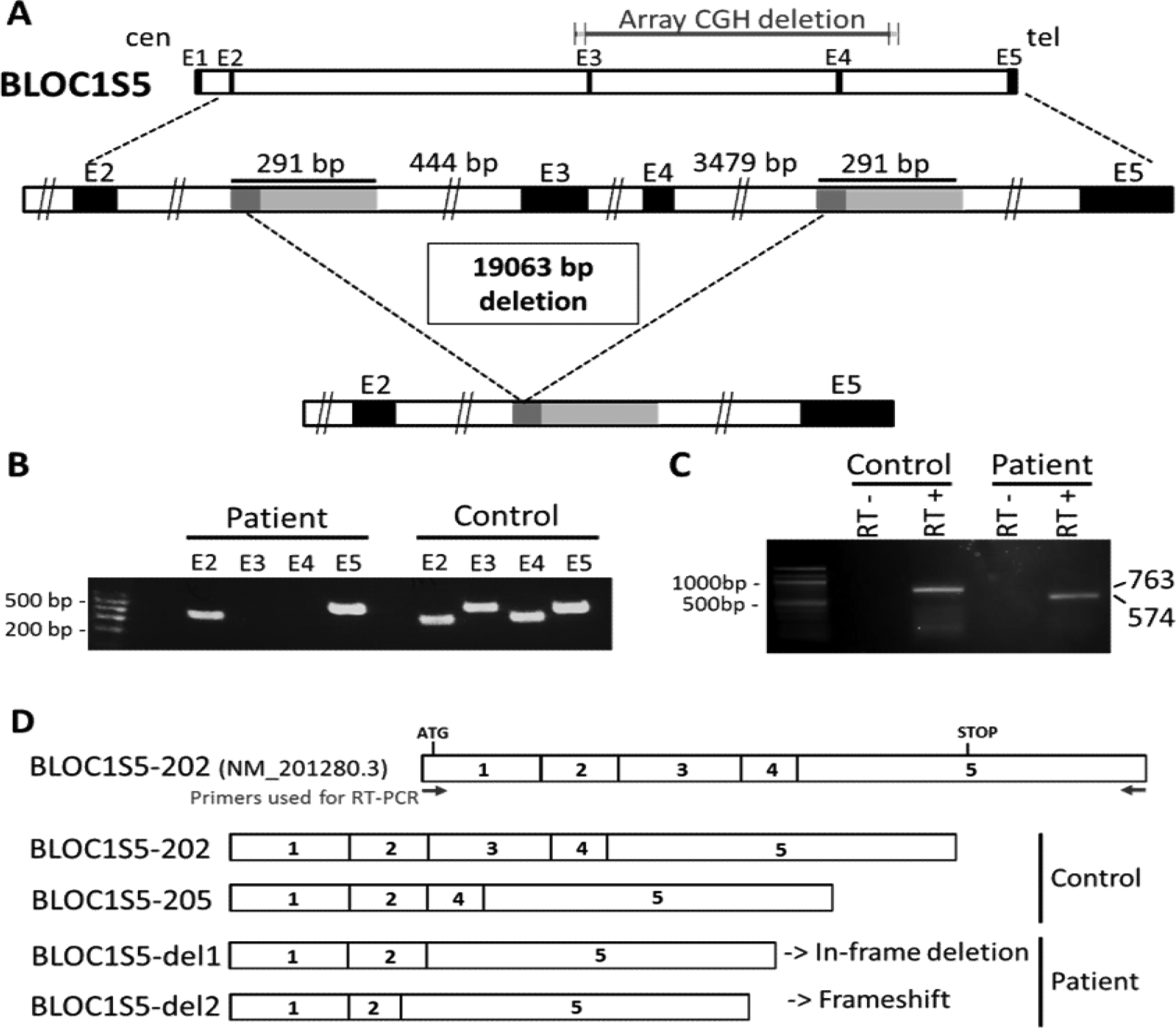
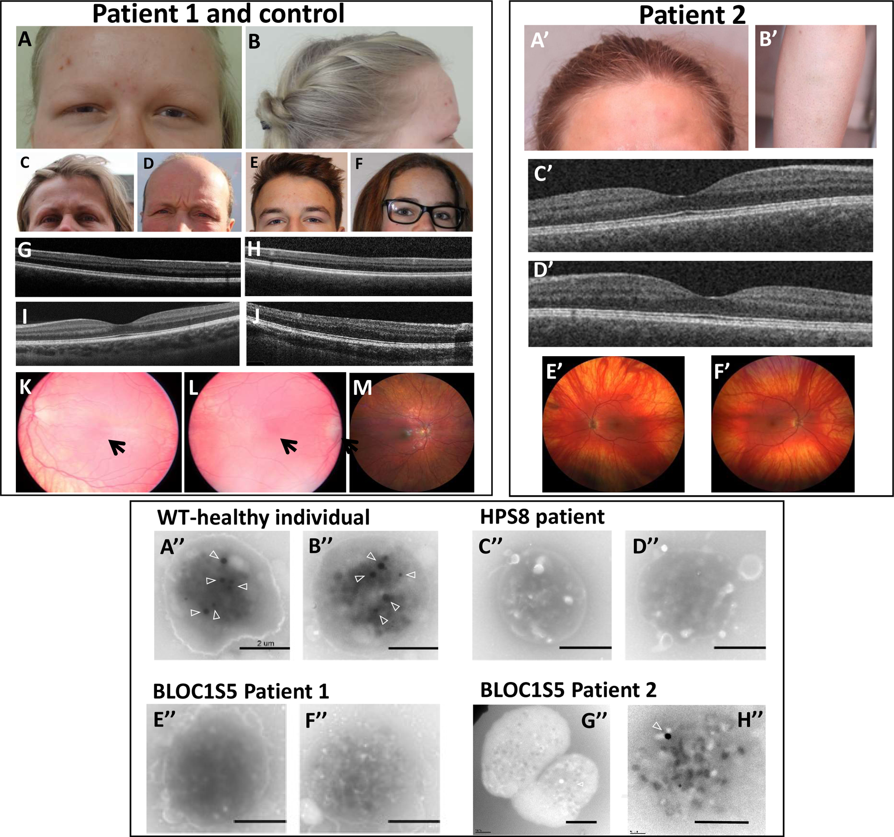
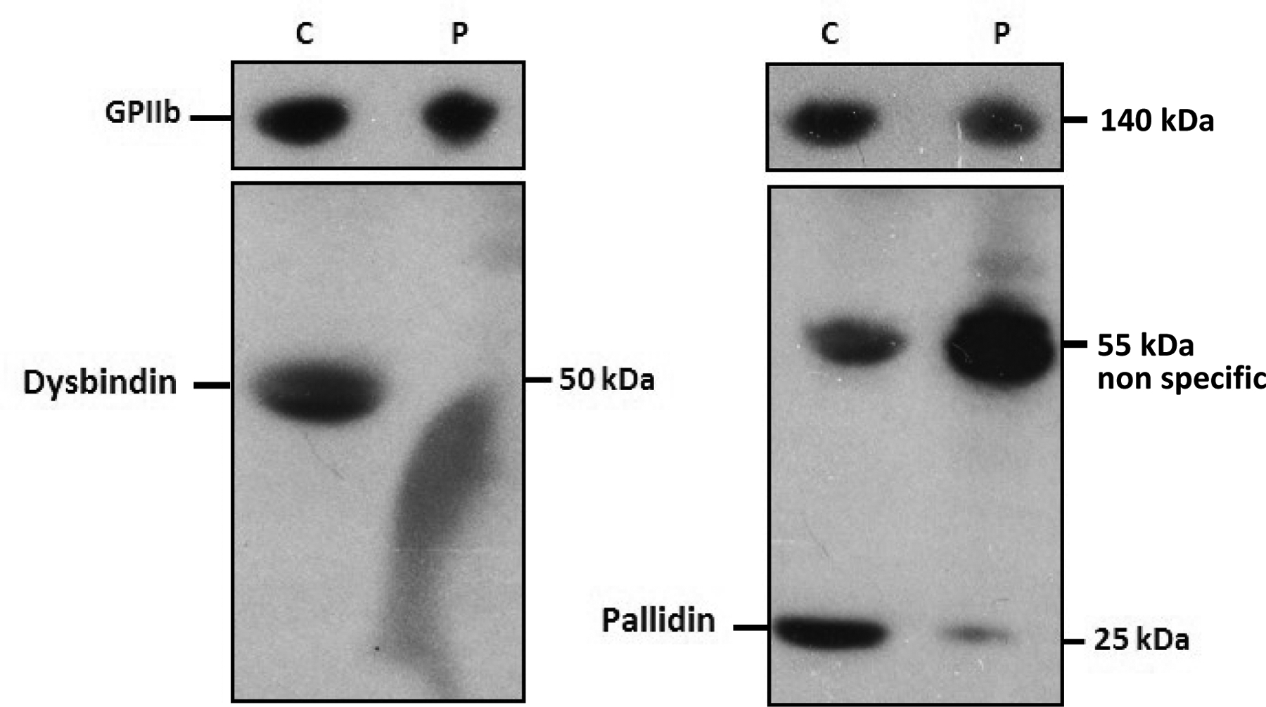
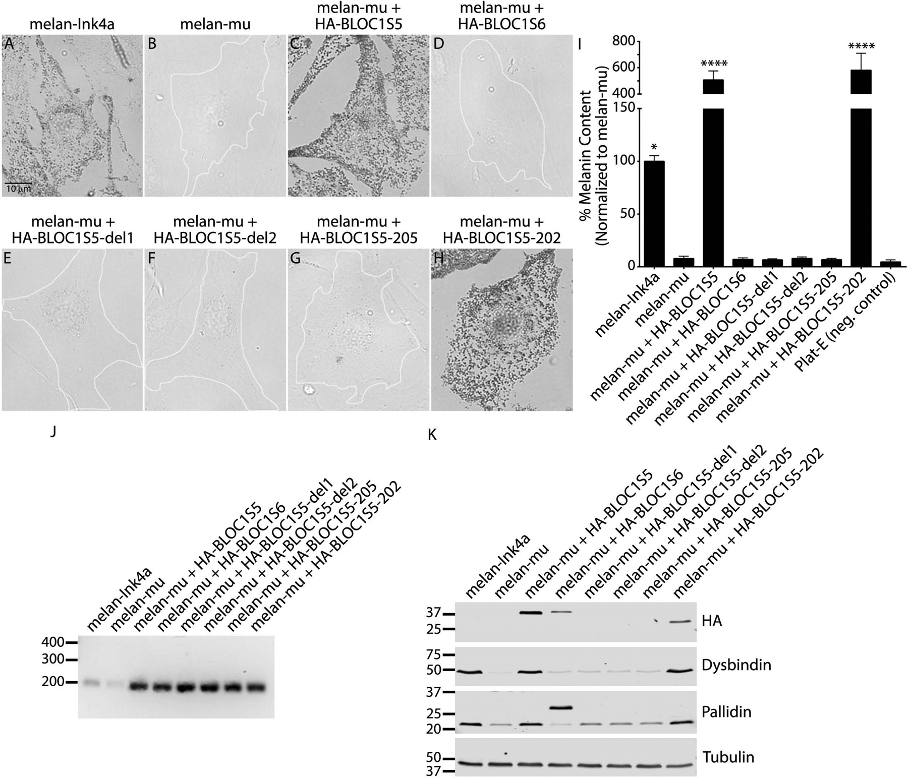
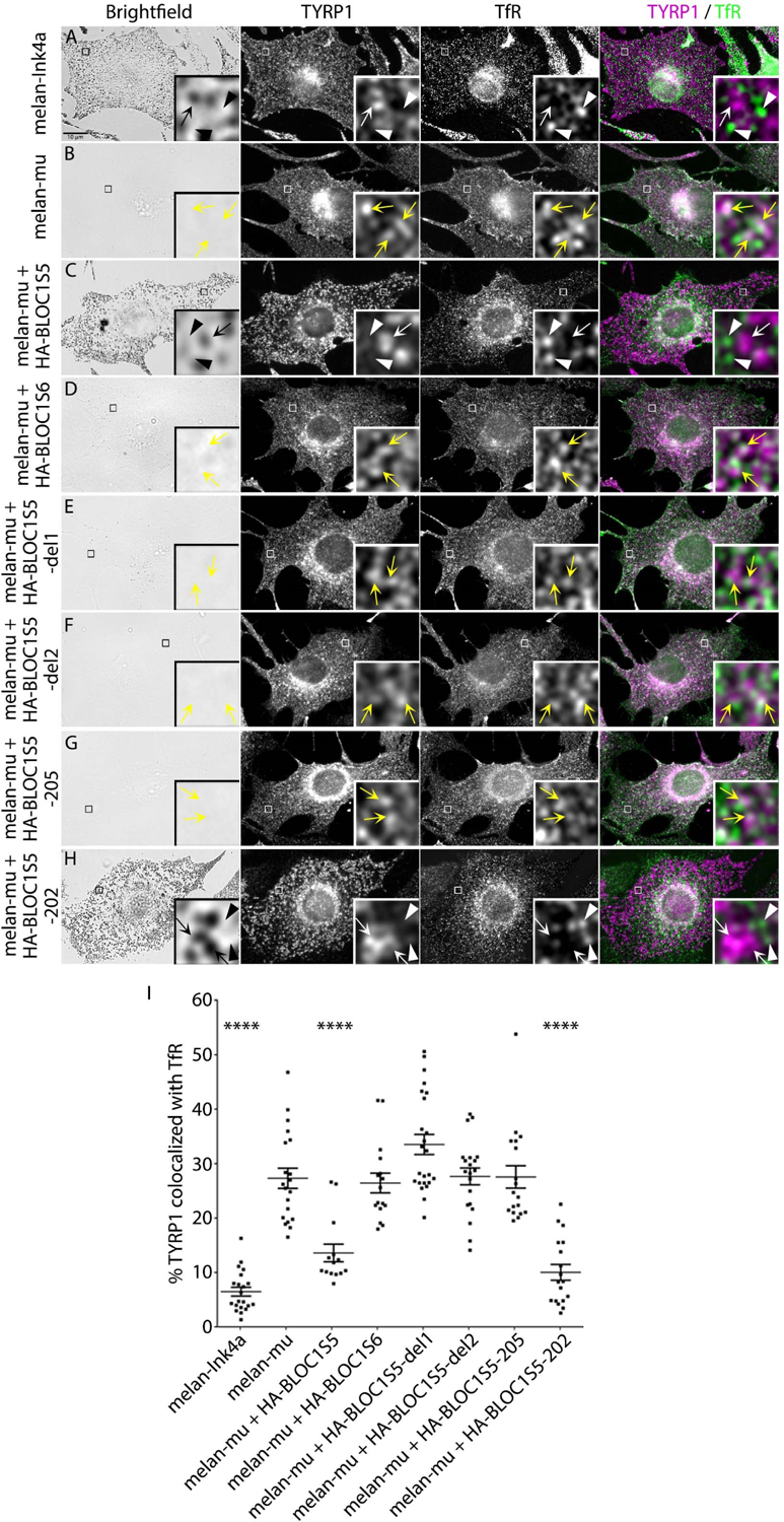
Results: We identified two unrelated patients with distinct homozygous variants of the BLOC1S5 gene. Patients had mild oculocutaneous albinism, moderate bleeding diathesis, platelet aggregation deficit, and a dramatically decreased number of platelet dense granules, all signs compatible with HPS. Functional tests performed on platelets of one patient displayed an absence of the obligate multisubunit complex BLOC-1, showing that the variant disrupts BLOC1S5 function and impairs BLOC-1 assembly. Expression of the patient-derived BLOC1S5 deletion in nonpigmented murine Bloc1s5-/- melan-mu melanocytes failed to rescue pigmentation, the assembly of a functional BLOC-1 complex, and melanosome cargo trafficking, unlike the wild-type allele.

Conclusion: Mutation of BLOC1S5 is disease-causing, and we propose that BLOC1S5 is the gene for a new form of Hermansky-Pudlak syndrome, HPS-11.

Keywords: BLOC-1; BLOC1S5; Hermansky–Pudlak syndrome; albinism; pathogenic variant.


Conflict of interest statement

Conflict of Interest Notification

The authors declare no conflict of interest.

Figures

Figure 1.
Figure 1.. Characterization of the BLOC1S5 deletion present in Patient 1.
A) Schematic representation of the deletion. Top: genomic representation of the BLOC1S5 gene (approximatively to scale). The five exons are represented as black boxes. cen: centromeric side; tel: telomeric side. Middle: The region spanning exons 2 – 5 of the BLOC1S5 gene is expanded. The light gray boxes represent the 291 bp highly homologous segments (see Supplementary Data for details). The dark grey boxes represent the 57 bp perfectly homologous segment where non-allelic homologous recombination appears to have taken place in Patient 1. Bottom: Deleted allele resulting from non-allelic homologous recombination, showing that the region between the two 291 bp highly homologous segments is missing. B) PCR amplification of exons 2, 3, 4 and 5 of BLOC1S5 in genomic DNA from a control individual and from Patient 1. A size marker (100 bp ladder) is shown on the left. Exons 3 and 4 are not amplified from the patient’s DNA. C) Reverse-transcription PCR from leukocytes’ RNA of a control individual and Patient 1. RT-PCR primers where derived from BLOC1S5 exons 1 and 5, as shown in D). RT- : without reverse transcriptase. RT+ : with reverse transcriptase. Bands at 763 bp (corresponding to cDNA BLOC1S5–202) and at 574 bp (corresponding to cDNA BLOC1S5-del1) are observed in the control and the patient, respectively. D) Schematic representation of the mRNA isoforms identified in the control individual and in Patient 1, as described in the main text. BLOC1S5–202 represents the theoretical wild-type isoform. Sizes of the complementary DNAs (cDNAs) corresponding to the different isoforms are BLOC1S5–202 (763bp) and BLOC1S5–205 (633bp), BLOC1S5-del1 (574bp), and BLOC1S5-del2 (542bp).
Figure 2.
Figure 2.. Clinical and platelet phenotype of Patients 1 and 2.
Upper left and right panel: cutaneous and ocular phenotype. Photographs show hypopigmentation of hair and skin of Patient 1 (A,B), compared to her unaffected mother (C), father (D), brother (E) and sister (F), and of Patient 2 (A’,B’). Bilateral foveal hypoplasia is seen in Patient 1 (G: left eye; H: right eye), compared to a normal control (I) and a positive control (OCA1 patient) (J). Patient 2 does not have foveal hypoplasia (C’: left eye; D’: right eye). Retinal hypopigmentation is seen in Patient 1 (K: left eye; L: right eye) compared to a normal control (M). Arrows point to hypoplasia of the macula. Patient 2 has low grade retinal hypopigmentation (E’: left eye; F’: right eye). Lower panel: whole mount electron microscopy of platelets. Platelets in healthy individuals displayed dense granules typically observed as round electron-dense structures (arrowheads) (A’’-B’’). Platelets from a HPS8 patient were used as a positive control for the absence of platelet dense granules (C’’-D’’). Platelets from the BLOC1S5 Patient 1 harbored no round electron-dense structures (E’’-F’’). Most platelets from the BLOC1S5 Patient 2 harbored no round electron-dense structures (G’’-H’’). Scale bar: 2 μm.
Figure 3.
Figure 3.. Western blots of platelet protein extracts from Patient 1 (P) and a healthy control (C).
Platelet lysates were analyzed with anti-Dysbindin (left), anti-Pallidin (right) antibodies. An anti-GPIIb antibody was used as a reference. Sizes of detected proteins are indicated in kilo-Dalton (kDa).
Figure 4.
Figure 4.. Patient 1-derived BLOC1S5 transgenes do not restore pigmentation to mouse Bloc1s5mu/mu melanocytes.
(a-h) Melanin content of fixed mouse melanocyte cell lines melan-Ink4a (wild-type), melan-mu (from Bloc1s5mu/mu mice) or melan-mu stably expressing the indicated HA.11-tagged Muted (BLOC1S5) or Pallidin (BLOC1S6) transgenes was assessed by bright field microscopy. Unpigmented cells are outlined in white. Scale, 10 μm. (i) Melanin content in lysates from the indicated cell lines was assayed by spectrophotometry. Plat-E is a non-pigmented cell line used as a negative control. Data are normalized to values from melan-mu and represent mean ± SEM from at least three experiments. Statistical analysis was performed using the Kruskal–Wallis test by ranks. *, P < 0.05; ****, P < 0.0001. (j) RT-PCR analysis to confirm transgene expression. cDNA was amplified from each of the indicated cell lines using primers for the hygromycin resistance gene expressed from the internal ribosome entry site of the pBMN-Hygro-IRES retroviral backbone, and products were fractionated by agarose gel electrophoresis. Shown is a representative of two experiments. Positions of DNA markers (bp) are shown at left. (k) Whole-cell lysates of indicated cell lines fractionated by SDS-PAGE were immunoblotted for the HA.11 epitope tag, BLOC-1 subunits Dysbindin and Pallidin, or γ-tubulin as a control. Shown is a representative of three experiments. Relevant bands (right) and positions of molecular weight markers (kDa, left) are indicated. Note, the higher molecular weight band in the Pallidin blot is HA-tagged Pallidin, whereas the lower molecular weight band represents endogenous Pallidin.
Figure 5.
Figure 5.. Patient 1-derived BLOC1S5 transgenes do not restore BLOC-1-dependent cargo transport in mouse Bloc1s5mu/mu melanocytes.
(a-h) Mouse melanocyte cell lines melan-Ink4a, melan-mu or melan-mu stably expressing the indicated HA.11-tagged Muted or Pallidin transgenes were fixed, immunolabeled for TYRP1 (magenta) and transferrin receptor (TfR, green), and analyzed by dIFM and by bright field microscopy to detect melanin. Scale, 10 μm. Insets of boxed regions are magnified ten times. TYRP1 localized on TfR-positive compartments (yellow arrowheads) or TYRP1 (white arrow) and TfR (white arrowhead) localized on discrete compartments are indicated. (i) Percent area of overlap between TYRP1 and TfR in melanocyte cell lines. Data are shown as dot plots with mean ± SEM from at least 14 cells representing three independent experiments. Statistical analysis was performed using a one-way ANOVA. ****, P < 0.0001.

Comment in

References

    1. Seward SL Jr, Gahl WA. Hermansky-Pudlak syndrome: health care throughout life. Pediatrics. 2013;132:153–160. - PubMed
    1. Delevoye C, Marks MS, Raposo G. Lysosome-related organelles as functional adaptations of the endolysosomal system. Curr Opin Cell Biol. 2019;59:147–158. - PMC - PubMed
    1. Bowman SL, Bi-Karchin J, Le L, Marks MS. The road to LROs: insights into lysosome-related organelles from Hermansky-Pudlak syndrome and other rare diseases. Traffic. 2019;20:404–435. - PMC - PubMed
    1. Nazarian R, Huizing M, Helip-Wooley A, Starcevic M, Gahl WA, Dell’Angelica EC. An immunoblotting assay to facilitate the molecular diagnosis of Hermansky-Pudlak syndrome. Mol Genet Metab. 2008;93:134–144. - PMC - PubMed
    1. Montoliu L, Marks MS. A new type of syndromic albinism associated with mutations in AP3D1. Pigment Cell Melanoma Res. 2017;30:5–7. - PMC - PubMed

Publication types