

The Effects of Sidt2 on the Inflammatory Pathway in Mouse Mesangial Cells
- PMID: 32565723
- PMCID: PMC7275211
- DOI: 10.1155/2020/3560793
The Effects of Sidt2 on the Inflammatory Pathway in Mouse Mesangial Cells
Abstract
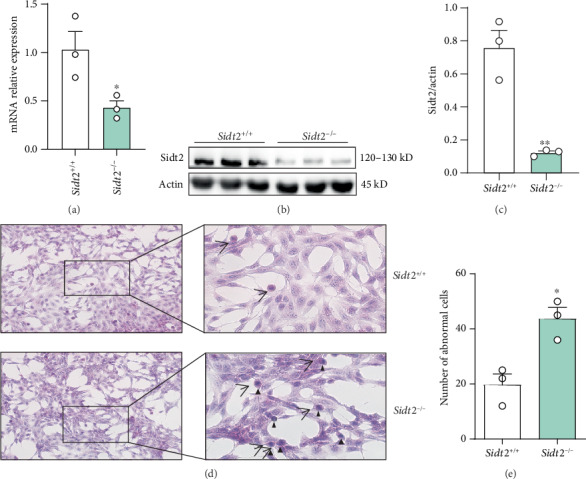
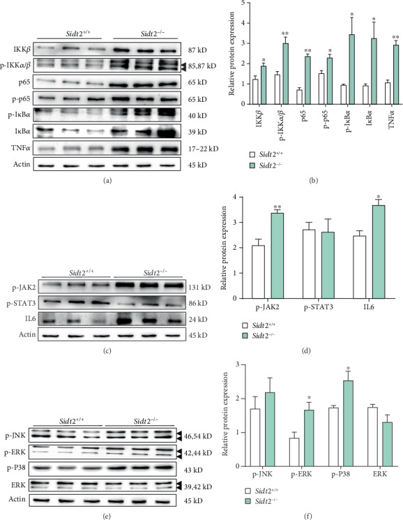
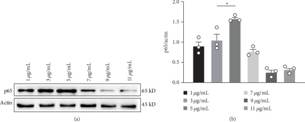
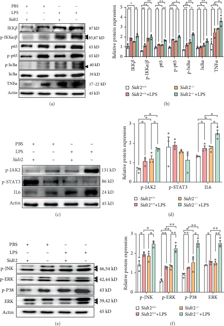
In patients with chronic kidney disease, the abnormal activation of inflammatory pathways is usually an important factor leading to renal fibrosis and further deterioration of renal function. Finding effective intervention targets of the inflammatory signaling pathway is an important way to treat chronic kidney disease. As a newly discovered lysosomal membrane protein, the correlation between SID1 transmembrane family member 2 (Sidt2) and the inflammatory signaling pathway has not been reported. The aim of this study was to investigate the effect of Sidt2 on inflammation by inhibiting the expression of the Sidt2 gene in a mouse mesangial cell line mediated by a lentiviral CRISPR/Cas9 vector. Hematoxylin and eosin staining and microscopy found that the mesangial cells lost their normal morphology after inhibiting the expression of Sidt2, showing that the cell body became smaller, the edge between the cells was unclear, and part of the nucleus was pyknotic and fragmented, appearing blue-black. The expressions of IKK β, p-IKK α/β, NF-κB p65, p-NF-κB p65, p-IκBα, IκBα, and TNF-α in the NF-κB pathway of the Sidt2 -/- group were higher than those of the Sidt2 +/+ group. p-Jak2 and IL6 increased in the Jak/Stat pathway, and p-ERK and p-P38 increased in the MAPK pathway. The expressions of IKK β, p-IKK α/β, NF-κB p65, p-NF-κB p65, p-IκBα, IκBα, and TNF-α in the NF-κB pathway of the Sidt2 +/++LPS group were significantly higher than those in the Sidt2 +/+ group. The expressions of IKK β, p-IKK α/β, NF-κB p65, p-NF-κB p65, p-IκBα, IκBα, and TNF-α in the Sidt2 -/-+LPS group were higher than those in the Sidt2 -/- group. The expressions of p-IKK α/β, NF-κB p65, p-NF-κB p65, p-IκBα, IκBα, and TNF-α in the Sidt2 -/-+LPS group were higher than those in the Sidt2 +/++LPS group. In the Jak/Stat pathway, the protein expressions of p-Jak2 and IL6 in the Sidt2 +/++LPS group were higher than those in the Sidt2 +/+ group. The expressions of p-Jak2 and IL6 in the Sidt2 -/-+LPS group were higher than those in the Sidt2 -/- group. The expressions of p-Jak2 and IL6 in the Sidt2 -/-+LPS group were higher than those in the Sidt2 +/++LPS group. The expressions of p-JNK, p-ERK, p-P38, and ERK in the MAPK pathway in the Sidt2 +/++LPS group were higher than those in the Sidt2 +/+ group. The expressions of p-JNK, p-ERK, p-P38, and ERK in the Sidt2 -/-+LPS group were higher than those in the Sidt2 -/- group. The expressions of p-JNK, p-ERK, p-P38, and ERK in the Sidt2 -/-+LPS group were higher than those in the Sidt2 +/++LPS group. These data suggested that deletion of the Sidt2 gene changed the three inflammatory signal pathways, eventually leading to the damage of glomerular mesangial cells in mice.
Copyright © 2020 Hui Sun et al.
Conflict of interest statement
The authors declare no conflicts of interest.
Figures
Similar articles
-
[Regulatory effects of lanthanum chloride on the activation of nuclear factor kappa B inhibitor kinase beta induced by tumor necrosis factor alpha].Zhonghua Shao Shang Za Zhi. 2013 Dec;29(6):531-6. Zhonghua Shao Shang Za Zhi. 2013. PMID: 24495640 Chinese.
-
Kaempferol attenuates inflammation in lipopolysaccharide-induced gallbladder epithelial cells by inhibiting the MAPK/NF-κB signaling pathway.Chem Biol Drug Des. 2024 Apr;103(4):e14519. doi: 10.1111/cbdd.14519. Chem Biol Drug Des. 2024. PMID: 38570708
-
TLR2 regulates allergic airway inflammation through NF-κB and MAPK signaling pathways in asthmatic mice.Eur Rev Med Pharmacol Sci. 2018 May;22(10):3138-3146. doi: 10.26355/eurrev_201805_15073. Eur Rev Med Pharmacol Sci. 2018. PMID: 29863259
-
IκB kinase regulation of the TPL-2/ERK MAPK pathway.Immunol Rev. 2012 Mar;246(1):168-82. doi: 10.1111/j.1600-065X.2012.01104.x. Immunol Rev. 2012. PMID: 22435554 Review.
-
Regulation of NF-κB by TNF family cytokines.Semin Immunol. 2014 Jun;26(3):253-66. doi: 10.1016/j.smim.2014.05.004. Epub 2014 Jun 21. Semin Immunol. 2014. PMID: 24958609 Free PMC article. Review.
Cited by
-
[Lysosomal membrane protein Sidt2 knockout induces apoptosis of human hepatocytes in vitro independent of the autophagy-lysosomal pathway].Nan Fang Yi Ke Da Xue Xue Bao. 2023 Apr 20;43(4):637-643. doi: 10.12122/j.issn.1673-4254.2023.04.18. Nan Fang Yi Ke Da Xue Xue Bao. 2023. PMID: 37202201 Free PMC article. Chinese.
-
CCL24 Protects Renal Function by Controlling Inflammation in Podocytes.Dis Markers. 2021 Jun 16;2021:8837825. doi: 10.1155/2021/8837825. eCollection 2021. Dis Markers. 2021. PMID: 34221188 Free PMC article.
-
Structural insight into the human SID1 transmembrane family member 2 reveals its lipid hydrolytic activity.Nat Commun. 2023 Jun 15;14(1):3568. doi: 10.1038/s41467-023-39335-2. Nat Commun. 2023. PMID: 37322007 Free PMC article.
-
Modified Gexiazhuyu decoction alleviates chronic salpingitis p38 signaling pathway.J Tradit Chin Med. 2022 Apr;42(2):213-220. doi: 10.19852/j.cnki.jtcm.2022.02.003. J Tradit Chin Med. 2022. PMID: 35473341 Free PMC article.
-
Sidt2 is a key protein in the autophagy-lysosomal degradation pathway and is essential for the maintenance of kidney structure and filtration function.Cell Death Dis. 2021 Dec 18;13(1):7. doi: 10.1038/s41419-021-04453-6. Cell Death Dis. 2021. PMID: 34923568 Free PMC article.
References
-
- Chen M. F., et al. Liou S. S., Hong T. Y., Kao S. T., Liu I. M. Gigantol has protective effects against high glucose-evoked nephrotoxicity in mouse glomerulus mesangial cells by suppressing ROS/MAPK/NF-κB signaling pathways. Molecules. 2019;24(1):p. 80. doi: 10.3390/molecules24010080. - DOI - PMC - PubMed
MeSH terms
Substances
LinkOut - more resources
Full Text Sources
Molecular Biology Databases
Research Materials
Miscellaneous
