. 2020 May 30:2020:6458791.
doi: 10.1155/2020/6458791. eCollection 2020.

hnRNPA2/B1 Ameliorates LPS-Induced Endothelial Injury through NF- κ B Pathway and VE-Cadherin/ β-Catenin Signaling Modulation In Vitro

Affiliations

hnRNPA2/B1 Ameliorates LPS-Induced Endothelial Injury through NF- κ B Pathway and VE-Cadherin/ β-Catenin Signaling Modulation In Vitro

Yi Chen et al. Mediators Inflamm. .

Abstract

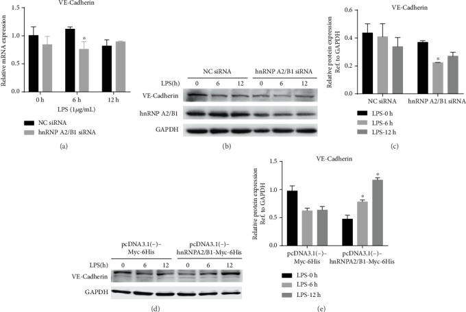
Heterogeneous nuclear ribonucleoprotein A2/B1 (hnRNPA2/B1) is a protein involved in the regulation of RNA processing, cell metabolism, migration, proliferation, and apoptosis. However, the effect of hnRNPA2/B1 on injured endothelial cells (ECs) remains unclear. We investigated the effect of hnRNPA2/B1 on lipopolysaccharide- (LPS-) induced vascular endothelial injury in human umbilical vein endothelial cells (HUVECs) and the underlying mechanisms. LPS was used to induce EC injury, and the roles of hnRNPA2/B1 in EC barrier dysfunction and inflammatory responses were measured by testing endothelial permeability and the expression of inflammatory factors after the suppression and overexpression of hnRNPA2/B1. To explore the underlying mechanism by which hnRNPA2/B1 regulates endothelial injury, we studied the VE-cadherin/β-catenin pathway and NF-κB activation in HUVECs. The results showed that hnRNPA2/B1 was elevated in LPS-stimulated HUVECs. Moreover, knockdown of hnRNPA2/B1 aggravated endothelial injury by increasing EC permeability and promoting the secretion of the inflammatory cytokines TNF-α, IL-1β, and IL-6. Overexpression of hnRNPA2/B1 can reduce the permeability and inflammatory response of HUVEC stimulated by LPS in vitro, while increasing the expression of VE-Cadherin and β-catenin. Furthermore, the suppression of hnRNPA2/B1 increased the LPS-induced NF-κB activation and reduced the VE-cadherin/β-catenin pathway. Taken together, these results suggest that hnRNPA2/B1 can regulate LPS-induced EC damage through regulating the NF-κB and VE-cadherin/β-catenin pathways.


Conflict of interest statement

The authors declare no conflict of interest.

Figures

Figure 1
Figure 1
hnRNPA2/B1 expression in LPS-stimulated HUVECs. HUVECs were treated with 1 μg/mL LPS for 0 h, 6 h, 12 h, and 24 h. (a) hnRNPA2/B1 mRNA expression levels were quantified by qRT-PCR, and β-actin was used as a normalization control. (b, c) hnRNPA2/B1 protein levels were measured by Western blotting, and GAPDH was used as an internal control. p < 0.05, ∗∗p < 0.01, ∗∗∗p < 0.001.
Figure 2
Figure 2
Effect of hnRNPA2/B1 on endothelial permeability in HUVECs. HUVECs were transfected with hnRNPA2/B1 siRNA/negative control (NC) siRNA or pcDNA3.1(-)-Myc-6His/pcDNA3.1(-)-hnRNPA2/B1-Myc-6His plasmid for 36 h and then stimulated with 1 μg/mL LPS for different lengths of time. The hnRNPA2/B1 knockdown efficiency was determined by qRT-PCR (a) and Western blotting (b, c). The hnRNPA2/B1 overexpression efficiency was determined by Western blotting (d, e). (f, h) TEER values were measured every 2 h for 24 h of LPS stimulation. (g, i) Endothelial permeability was determined by FITC-dextran flux analysis 12 h after LPS administration. Data are expressed as mean ± SD (n = 3), vs. Negative control (siRNA/plasmid) p < 0.05, # vs. Negative control (siRNA/plasmid) + LPSp < 0.05.
Figure 3
Figure 3
Effect of hnRNPA2/B1 on inflammatory cytokine expression in HUVECs. Twelve hours after LPS treatment, HUVECs and culture supernatant were collected. (a, c) The expression levels of IL-1β, TNF-α, and IL-6 were measured by RT-qPCR. β-Actin was used as a normalization control. (b, d) IL-1β, TNF-α, and IL-6 concentrations were assayed by ELISA. Data are expressed as mean ± SD (n = 3), vs. Negative control (siRNA/plasmid) p < 0.05.
Figure 4
Figure 4
Effect of hnRNPA2/B1 on the expression of adherens junction molecules in LPS-stimulated HUVECs. HUVECs were transfected with hnRNPA2/B1 siRNA/negative control (NC) siRNA or pcDNA3.1(-)-Myc-6His/pcDNA3.1(-)-hnRNPA2/B1-Myc-6His plasmid for 36 h and then stimulated with 1 μg/mL LPS for 0, 6, and 12 h. The mRNA and protein were extracted, and VE-cadherin and GAPDH mRNA and protein levels were detected by RT-qPCR and Western blotting, respectively. Data are expressed as mean ± SD (n = 3), vs. Negative control (siRNA/plasmid) p < 0.05.
Figure 5
Figure 5
The effect of hnRNPA2/B1 reverses the LPS-induced downregulation of β-catenin and endothelial hyperpermeability in HUVECs. Twelve hours after LPS treatment, HUVECs, and culture supernatant were collected. (a) The mRNA expression level of β-catenin was measured by RT-qPCR. β-Actin was used as a normalization control. (b, c, e, f) The protein expression level of β-catenin was detected by Western blotting. (d) Immunofluorescent staining for β-catenin (green) in HUVECs stained with DAPI (blue). Arrows indicate areas of cell-cell adhesion. Scale bars, 50 μm. Data are expressed as mean ± SD (n = 3), vs. Negative control (siRNA/plasmid) p < 0.05.
Figure 6
Figure 6
Effect of hnRNPA2/B1 knockdown on LPS-induced NF-κB signaling activation. HUVECs transfected with siRNA (hnRNPA2/B1 or negative control) were cultured for 36 h and then treated with 1 μg/mL LPS for 0, 6, and 12 h. (a, b) NF-κB phosphorylation was examined by Western blotting, and GAPDH was used as an internal reference. Data are expressed as mean ± SD (n = 3), vs. NC siRNA p < 0.05.

References

    1. Singer M., Deutschman C. S., Seymour C. W., et al. The Third International Consensus Definitions for Sepsis and Septic Shock (Sepsis-3) JAMA. 2016;315(8):801–810. doi: 10.1001/jama.2016.0287. - DOI - PMC - PubMed
    1. Cecconi M., Evans L., Levy M., Rhodes A. Sepsis and septic shock. The Lancet. 2018;392(10141):75–87. doi: 10.1016/S0140-6736(18)30696-2. - DOI - PubMed
    1. Dellinger R. P., Levy M. M., Rhodes A., et al. Surviving Sepsis Campaign: international guidelines for management of severe sepsis and septic shock, 2012. Intensive Care Medicine. 2013;39(2):165–228. doi: 10.1007/s00134-012-2769-8. - DOI - PMC - PubMed
    1. Seymour C. W., Rosengart M. R. Septic Shock. JAMA. 2015;314(7):708–717. doi: 10.1001/jama.2015.7885. - DOI - PMC - PubMed
    1. Ait-Oufella H., Maury E., Lehoux S., Guidet B., Offenstadt G. The endothelium: physiological functions and role in microcirculatory failure during severe sepsis. Intensive Care Medicine. 2010;36(8):1286–1298. doi: 10.1007/s00134-010-1893-6. - DOI - PubMed

LinkOut - more resources