Review
. 2020 May 28:2020:4649081.
doi: 10.1155/2020/4649081. eCollection 2020.

Thoracic Complications in Behçet's Disease: Imaging Findings

Affiliations
Review

Thoracic Complications in Behçet's Disease: Imaging Findings

Kemal Ödev et al. Can Respir J. .

Abstract

Behçet's disease (BD) causes vascular inflammation and necrosis in a wide range of organs and tissues. In the thorax, it may cause vascular complications, affecting the aorta, brachiocephalic arteries, bronchial arteries, pulmonary arteries, pulmonary veins, capillaries, and mediastinal and thoracic inlet veins. In BD, chest radiograph is commonly used for the initial assessment of pulmonary symptoms and complications and for follow-up and establishment of the response to treatment. With the advancement of helical or multislice computed tomography (CT) technologies, such noninvasive imaging techniques have been employed for the diagnosis of vascular lesions, vascular complications, and pulmonary parenchymal manifestations of BD. CT scan (especially, CT angiography) has been used to determine the presence and severity of pulmonary complications without resorting to more invasive procedures, in conjunction with gadolinium-enhanced three-dimensional (3D) gradient-echo magnetic resonance (MR) imaging with the subtraction of arterial phase images. These radiologic methods have characteristics that are complementary to each other in diagnosis of the thoracic complications in BD. 3D ultrashort echo time (UTE) MR imaging (MRI) could potentially yield superior image quality for pulmonary vessels and lung parenchyma when compared with breath-hold 3D MR angiography.


Conflict of interest statement

The authors of this manuscript declare no relationships with any companies whose products or services may be related to the subject matter of the article.

Figures
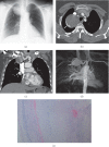
Figure 1
Figure 1
A 45-year-old woman with chronic cough, dyspnea, and hemoptysis. She had no family history of Behçet's disease or familial vascular disorder. (a) Chest radiograph showing a focal right paratracheal mass (white arrow). (b) Axial contrast-enhanced chest CT scan (mediastinal window) at the level of the thoracic inlet showing a mass with curvilinear calcification (white arrow). (c) Coronal contrast-enhanced chest CT scan (mediastinal window) showing a round mass (white arrow) in contact with the right subclavian artery (arrow head). Curvilinear calcification is visible in the wall of the mass. (d) Coronal maximum-intensity projection magnetic resonance angiography image showing a saccular aneurysm at the right subclavian artery (white arrow). The diagnosis of Behçet's disease was made after histopathological examination of the resected specimen following surgery. (e) Histopathology of resected subclavian arterial aneurysm. Hematoxylin-eosin, magnification x40. Chronic inflammatory process containing lymphocyte infiltration was detected in the adventitia that was compatible with diagnosis of Behçet's disease.
Figure 2
Figure 2
A 40-year-old woman with pulmonary artery aneurysm who had hemoptysis and chest pain. (a) Initial chest radiograph showing a round opacity at the inferior portion of the hilus on the right (arrow). (b) Axial contrast-enhanced chest CT scan (curved multiplanar reconstruction) (mediastinal window) showing a saccular aneurysm of the right interlobar artery with a circumferential mural thrombus (arrow).
Figure 3
Figure 3
Pulmonary artery aneurysm in a 37-year-old male with hemoptysis. (a) Axial contrast-enhanced CT scan (curved multiplanar reconstruction) (mediastinal window) showing an aneurysm of the right interlobar pulmonary artery (arrow). (b) MIP-reconstructed MRA image demonstrates an aneurysm of the right interlobar pulmonary artery aneurysm (long arrow).
Figure 4
Figure 4
A 40-year-old man with pulmonary artery aneurysms who had recurrent hemoptysis and chest pain and dyspnea. (a) Before treatment with immunosuppressive axial contrast-enhanced chest CT scan (mediastinal window) showing an aneurysm with lobulated contour on the right interlobar pulmonary artery (arrows). (b) Follow-up coronal maximum-intensity projection CT image showing multiple intraluminal filling defects within the upper lobe segmental branches of the right pulmonary artery (short arrows) and the right pulmonary artery with lobulated contour (arrow). (c) An axial contrast-enhanced CT scan through the lower chest showing basal segmental pulmonary arteries with central filling defects consistent with thromboembolism (arrows). Note. In this patient, deep vein thrombophlebitis and deep vein thrombosis in the lower extremities were not observed by Doppler ultrasonography. Thus, pulmonary thromboemboli probably developed as a complication of anticoagulant therapy. (d) Three years after treatment with immunosuppressive, axial maximum-intensity projection CT (curved multiplanar reconstruction) image shows regression of aneurysm on the right interlobar pulmonary artery (arrow heads). (e) High-resolution CT scan obtained 1-year later following immunosuppressive treatment showing a soft tissue nodule with air-crescent sign (arrow) within a cavity in the right upper lung and mycetoma showing a characteristic crescent of air between the mycetoma and the cavity wall. Radiological and serological tests confirmed Aspergillus fumigatus.
Figure 5
Figure 5
25-year-old man with multiple pulmonary artery aneurysms who had hemoptysis and chest pain with known BD. (a) Coronal maximum-intensity projection CT (curved multiplanar reconstruction) image showing multiple bilateral pulmonary artery aneurysms and mural thrombi (arrows). (b) Large right pulmonary artery aneurysm (long arrow) is partially lined by thrombus and densely opacified on a volumetric high-resolution CT. Lung window scan at the level (Figure 5(a)) shows the right pulmonary artery aneurysm with circumferential ground glass opacification because of surrounding haemorrhage (arrow): this finding was attributed to intraparenchymal haemorrhage. (c) Chest CT scans (lung window) obtained at different levels demonstrate multiple subpleural nodular infiltrations in the periphery of the right and left lung (arrows).
Figure 6
Figure 6
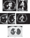
A 30-year-old male who presented with a history symptoms and signs of SVC syndrome for five years. He presented with a marked respiratory distress with cough and dyspnea. (a) Upper extremity contrast-enhanced venography shows bilateral occlusion of brachiocephalic veins and SVC. Also note the collateral veins in the chest wall and axillary region. (b) Follow-up axial contrast-enhanced chest CT scan shows nonopacification of the SVC (short arrow) and opacification of the multiple mediastinal veins (arrows). (c) Coronal maximum-intensity projection (MIP) reformation shows occlusion of the SVC due to thrombosis (long arrow) and bilaterally complete occlusion of the brachiocephalic, subclavian, and axillary veins. (d) Also, sagittal reformatted CT scan reveals that venous drainage from thorax is via the azygos system (arrow head) to SVC (arrow). (e) Coronal MIP reformation shows collateral flow through dilated pericardial vein (arrow head). (f) Coronal maximum-intensity (MIP) projection image from gadolinium-enhanced 3D MR angiography shows occlusion of the thoracic veins and SVC and dilated azygos vein (AV) and collateral veins at thoracic inlet level and chest wall (arrow heads) on the right (IVC: arrowhead). Behçet's disease was clinically diagnosed because of recurrent oral and genital ulcerations and positive pathergy test. The diagnosis was confirmed by histopathologic examination.
Figure 7
Figure 7
Superior vena cava syndrome in a 50-year-old man who had BD for ten years. (a) Before treatment with immunosuppressive, axial contrast-enhanced chest CT scan (mediastinal window) showing occlusion of innominate veins and SVC (thick arrow). (b) Sagittal oblique reformatted CT scan showing complete occlusion of the SVC (thin arrow) and dilated azygos vein (thick arrows). (c) Gadolinium-enhanced 3D MR venography showing bilaterally complete occlusion of major bilaterally brachiocephalic veins and SVC (long arrow) due to thrombosis and extensive collateral veins (short arrows) in the chest wall. (d) Coronal maximum-intensity images from 3D gadolinium-enhanced subtraction MR venogram obtained by subtracting from the arterial phase showing complete occlusion of brachiocephalic, subclavian, and axillar veins as well as SVC and extensive collateral circulation at chest wall on the left (black arrows). (e) After treatment with immunosuppressive, axial noncontrast 3D UTE-MRI shows SVC stenosis (arrow) and wall thickening of SVC (arrow). (f) Follow-up contrast-enhanced coronal MRA shows lack of opacification of SVC (arrows) and occlusion of bilaterally innominate veins and axillary veins after treatment with immunosuppressive.
Figure 8
Figure 8
Intracardiac thrombus. A contrast-enhanced chest CT scan demonstrates a low attenuating mass-like lesion (white arrow) in the left atrium without enhancement, pathologically proven an organized clot.

References

    1. Cadman E. C., Lundberg W. B., Mitchell M. S. Pulmonary manifestations in Behçet syndrome. Archives of Internal Medicine. 1976;136(8):944–947. doi: 10.1001/archinte.1976.03630080078023. - DOI - PubMed
    1. Chamberlain M. A. Behcet’s syndrome in 32 patients in Yorkshire. Annals of the Rheumatic Diseases. 1977;36(6):491–499. doi: 10.1136/ard.36.6.491. - DOI - PMC - PubMed
    1. Efthimiou J., Johnston C., Spiro S. G., et al. Pulmonary disease in behçet’s syndrome. QJM: An International Journal of Medicine. 1986;58:259–280. - PubMed
    1. Matsumoto T., Uekusa T., Fukuda Y. Vasculo-Behçet’s disease: a pathologic study of eight cases. Human Pathology. 1991;22(1):45–51. doi: 10.1016/0046-8177(91)90060-3. - DOI - PubMed
    1. Behçet H. Uber rezidivierende, aphthose, durchein Virus verusachte Gaschwure am Mund, am Auge und an den Genitalien. Dermatol Wochenschr. 1937;105:1152–1157.

MeSH terms