. 2020 May 20:2020:3245483.
doi: 10.1155/2020/3245483. eCollection 2020.

Wenxin Granule Ameliorates Hypoxia/Reoxygenation-Induced Oxidative Stress in Mitochondria via the PKC- δ/NOX2/ROS Pathway in H9c2 Cells

Affiliations

Wenxin Granule Ameliorates Hypoxia/Reoxygenation-Induced Oxidative Stress in Mitochondria via the PKC- δ/NOX2/ROS Pathway in H9c2 Cells

Qihui Jin et al. Oxid Med Cell Longev. .

Abstract

Myocardial infarction and following reperfusion therapy-induced myocardial ischemia/reperfusion (I/R) injury have been recognized as an important subject of cardiovascular disease with high mortality. As the antiarrhythmic agent, Wenxin Granule (WXG) is widely used to arrhythmia and heart failure. In our pilot study, we found the antioxidative potential of WXG in the treatment of myocardial I/R. This study is aimed at investigating whether WXG could treat cardiomyocyte hypoxia/reoxygenation (H/R) injury by inhibiting oxidative stress in mitochondria. The H9c2 cardiomyocyte cell line was subject to H/R stimuli to mimic I/R injury in vitro. WXG was added to the culture medium 24 h before H/R exposing as pretreatment. Protein kinase C-δ (PKC-δ) inhibitor rottlerin or PKC-δ lentivirus vectors were conducted on H9c2 cells to downregulate or overexpress PKC-δ protein. Then, the cell viability, oxidative stress levels, intracellular and mitochondrial ROS levels, mitochondrial function, and apoptosis index were analyzed. In addition, PKC-δ protein expression in each group was verified by western blot analysis. Compared with the control group, the PKC-δ protein level was significantly increased in the H/R group, which was remarkably improved by WXG or rottlerin. PKC-δ lentivirus vector-mediated PKC-δ overexpression was not reduced by WXG. WXG significantly improved H/R-induced cell injury, lower levels of SOD and GSH/GSSG ratio, higher levels of MDA, intracellular and mitochondrial ROS content, mitochondrial membrane potential and ATP loss, mitochondrial permeability transition pore opening, NOX2 activation, cytochrome C release, Bax/Bcl-2 ratio and cleaved caspase-3 increasing, and cell apoptosis. Similar findings were obtained from rottlerin treatment. However, the protective effects of WXG were abolished by PKC-δ overexpression, indicating that PKC-δ was a potential target of WXG treatment. Our findings demonstrated a novel mechanism by which WXG attenuated oxidative stress and mitochondrial dysfunction of H9c2 cells induced by H/R stimulation via inhibitory regulation of PKC-δ/NOX2/ROS signaling.


Conflict of interest statement

There is no conflict of interest in this article.

Figures

Figure 1
Figure 1
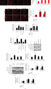
Overexpression of PKC-δ reversed the positive effect of WXG on H9c2 cells under H/R. (a, b) CCK-8 assay showed the proliferation of H9c2 cells under H/R (n = 6). (c, d) LDH release assay showed cell injury in each group (n = 6). (e–h) Western blot revealed PKC-δ protein expression (n = 3). Representative immunoblots were normalized to β-actin. ##P < 0.01, ###P < 0.001 vs. the control group; P < 0.05, ∗∗P < 0.01, and ∗∗∗P < 0.001 vs. the H/R group. &&P < 0.01, §§P < 0.01.
Figure 2
Figure 2
WXG rescued H/R-induced H9c2 cell oxidative stress injury which was reversed by PKC-δ overexpression. (a, b) SOD activity was determined by the SOD assay. (c, d) MDA content was estimated by the MDA assay. (e, f) GSH/GSSG ratios were estimated by the GSH and GSSG assay. N = 6 in each group. ##P < 0.01, ###P < 0.001 vs. the control group; P < 0.05, ∗∗P < 0.01, and ∗∗∗P < 0.001 vs. the H/R group. §§P < 0.01, §§§P < 0.001.
Figure 3
Figure 3
WXG suppressed ROS generation in H/R-induced H9c2 cells which was reversed by PKC-δ overexpression. (a–d) Representative fluorescence images (left panel) and quantification (right panel) of DHE fluorescence (n = 6). Sale bar =20 μM. (e, f) Quantification of MitoSOX fluorescence in each group (n = 6). (g) Quantification of DCF fluorescence in each group (n = 6). (h–m) Western blot revealed p47phxo (membrane/cytosolic) and gp91phox protein expression (n = 3). Representative immunoblots were normalized to β-actin. #P < 0.05, ##P < 0.01, and ###P < 0.001 vs. the control group; P < 0.05, ∗∗P < 0.01 vs. the H/R group. §P < 0.05, §§P < 0.01, and §§§P < 0.001.
Figure 4
Figure 4
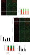
WXG inhibited the collapse of mitochondrial transmembrane potential and ATP level in H/R-induced H9c2 cells which was reversed by PKC-δ overexpression. (a, d) Representative fluorescence images of JC-1 fluorescence. Scale bar =20 μM. (b, e) Quantification (right panel) of JC-1 fluorescence (n = 6). (c, f) ATP level was assessed using an ATP detection kit (n = 6). ###P < 0.001 vs. the control group; P < 0.05, ∗∗P < 0.01 vs. the H/R group. §§§P < 0.001.
Figure 5
Figure 5
WXG decreased opening of the mitochondrial permeability transition pore in H/R-induced H9c2 cells which was reversed by PKC-δ overexpression. (a–d) Representative fluorescence images and quantification of calcein fluorescence (n = 6). Scale bar =50 μM. ###P < 0.001 vs. the control group; ∗∗∗P < 0.001 vs. the H/R group. §§§P < 0.001. (e–h) Western blot revealed cytochrome C protein expression (n = 3). Representative immunoblots were normalized to β-actin. ##P < 0.01, ###P < 0.001 vs. the control group; P < 0.05, ∗∗P < 0.01, and ∗∗∗P < 0.001 vs. the H/R group. §P < 0.05, §§§P < 0.001.
Figure 6
Figure 6
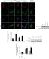
WXG decreased cytochrome C increase and release in H/R-induced H9c2 cells which was reversed by PKC-δ overexpression. (a) Representative fluorescence images of MitoTracker and cytochrome C fluorescence (n = 6). Scale bar = 5 μM. (b–e) Western blot revealed cytochrome C protein expression (n = 3). Representative immunoblots were normalized to β-actin. ##P < 0.01, ###P < 0.001 vs. the control group; P < 0.05, ∗∗P < 0.01 vs. the H/R group. §P < 0.05, §§§P < 0.001.
Figure 7
Figure 7
WXG reduced H/R-triggered apoptosis in H9c2 cells which was reversed by PKC-δ overexpression. (a–d) Representative fluorescence images of TUNEL (green) and DAPI (blue) and quantification of TUNEL-positive cells (n = 6). Scale bar = 100 μM. ###P < 0.001 vs. the control group; P < 0.05 vs. the H/R group. §P < 0.05.
Figure 8
Figure 8
WXG downregulated apoptosis protein in H/R-induced H9c2 cells which was reversed by PKC-δ overexpression. (a–f) Western blot revealed Bcl-2, Bax, and cleaved caspase-3 protein expression (n = 3). Representative immunoblots were normalized to β-actin. #P < 0.05, ##P < 0.01, and ###P < 0.001 vs. the control group; P < 0.05 vs. the H/R group. §P < 0.05.
Figure 9
Figure 9
WXG rescued H9c2 cells under H/R stimulation by attenuating mitochondrial oxidative stress via PKC-δ/NOX2/ROS inactivation.

References

    1. Liu J., Sui H., Zhao J., Wang Y. Osmotin protects H9c2 cells from simulated ischemia-reperfusion injury through AdipoR1/PI3K/AKT signaling pathway. Frontiers in physiology. 2017;8:p. 611. doi: 10.3389/fphys.2017.00611. - DOI - PMC - PubMed
    1. Chen W. R., Hu S. Y., Chen Y. D., et al. Effects of liraglutide on left ventricular function in patients with ST- segment elevation myocardial infarction undergoing primary percutaneous coronary intervention. American Heart Journal. 2015;170(5):845–854. doi: 10.1016/j.ahj.2015.07.014. - DOI - PubMed
    1. Heusch G., Musiolik J., Gedik N., Skyschally A. Mitochondrial STAT3 activation and cardioprotection by ischemic postconditioning in pigs with regional myocardial ischemia/reperfusion. Circulation Research. 2011;109(11):1302–1308. doi: 10.1161/CIRCRESAHA.111.255604. - DOI - PubMed
    1. Jin Q., Li R., Hu N., et al. DUSP1 alleviates cardiac ischemia/reperfusion injury by suppressing the Mff- required mitochondrial fission and Bnip3-related mitophagy via the JNK pathways. Redox Biology. 2018;14:576–587. doi: 10.1016/j.redox.2017.11.004. - DOI - PMC - PubMed
    1. Neri M., Riezzo I., Pascale N., Pomara C., Turillazzi E. Ischemia/reperfusion injury following acute myocardial infarction: a critical issue for clinicians and forensic pathologists. Mediators of inflammation. 2017;2017:14. doi: 10.1155/2017/7018393.7018393 - DOI - PMC - PubMed

MeSH terms