. 2020 May 23:2020:4951820.
doi: 10.1155/2020/4951820. eCollection 2020.

Huangbai Liniment Accelerated Wound Healing by Activating Nrf2 Signaling in Diabetes

Affiliations

Huangbai Liniment Accelerated Wound Healing by Activating Nrf2 Signaling in Diabetes

Jingjing Zhang et al. Oxid Med Cell Longev. .

Abstract

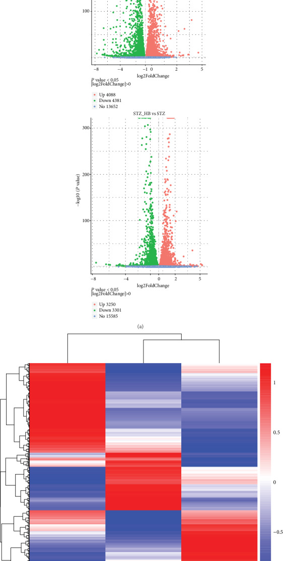
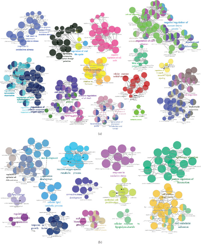
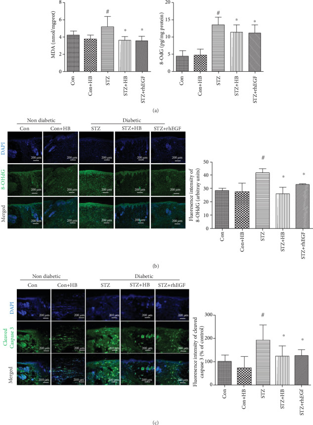
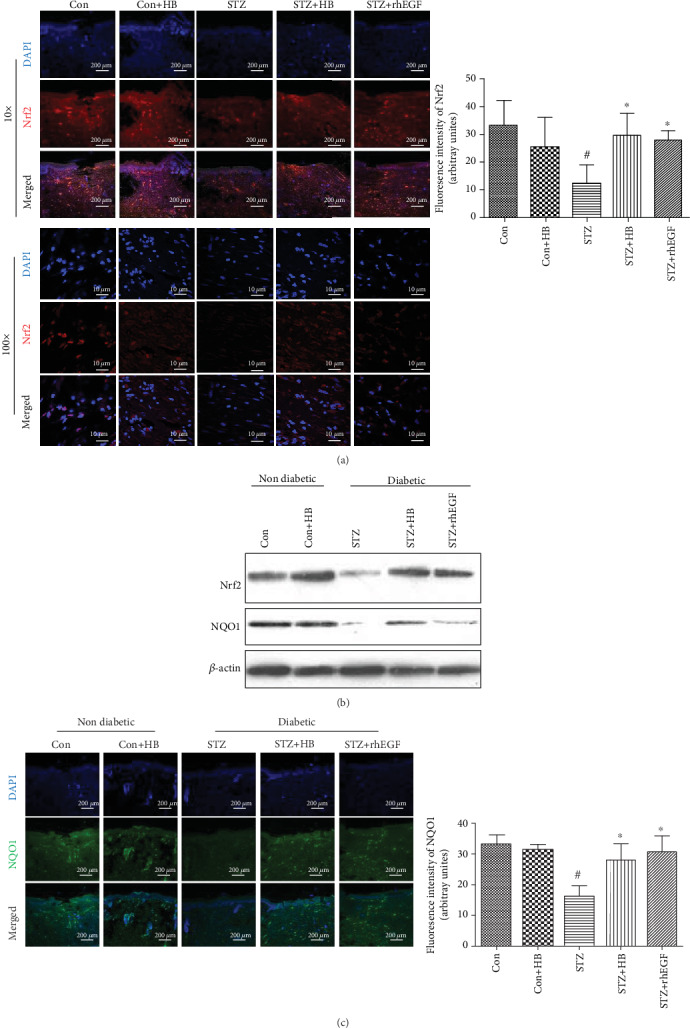
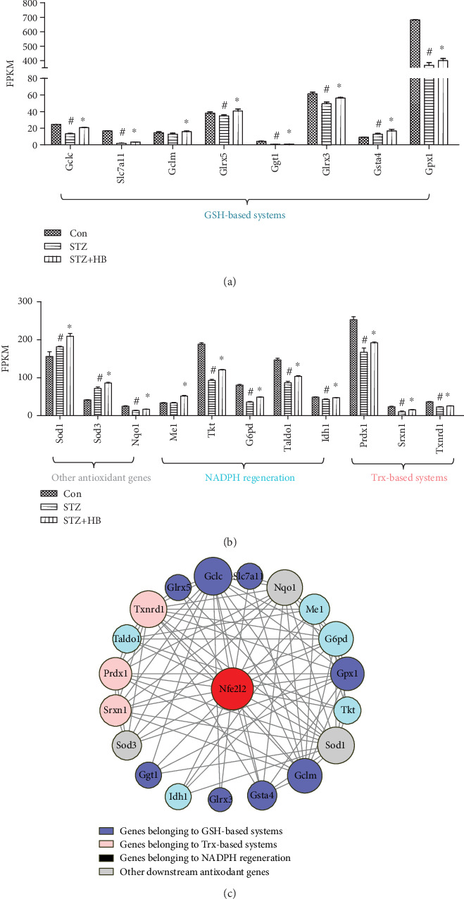
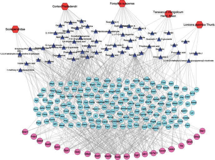
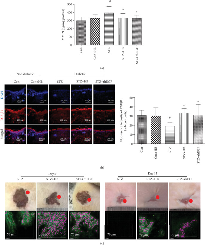
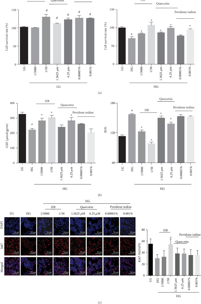
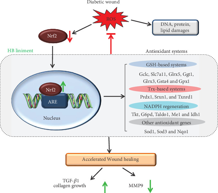
As a serious complication of diabetes, nonhealing skin ulcer leads to high mortality and disability in diabetic patients. However, limited therapy is available in managing diabetic wounds. In this study, RNA-seq technology was used to systematically investigate the effect of Huangbai (HB) liniment, a traditional Chinese medicine, on the streptozotocin- (STZ-) induced diabetic wound. HB liniment significantly accelerated the wound closure and enhanced the generation of extracellular matrix in diabetic rats, and oxidative stress was identified to play a vital role in HB-mediated wound healing. Importantly, HB liniment activated nuclear factor erythroid-derived 2-like 2 (Nrf2) and its downstream antioxidant genes (e.g., genes involved in glutathione system, thioredoxin system, and GAPDH generation as well as other antioxidant genes), which inhibited oxidative damage and apoptosis. By associating drug targets of HB liniment with Nrf2 and its downstream genes, 54 components in HB liniment were screened out, and the majority was from Cortex Phellodendri and Forsythia suspensa. Additionally, HB liniment enhanced TGF-β1 and reduced MMP9 level, accelerating wound healing in diabetes. The in vitro experiment showed HB facilitated cell proliferation and inhibited oxidative damage in high glucose-induced HaCaT cells. Our findings provided the experimental evidence for the treatment of diabetic wound with HB, clarified the potential mechanism of HB, and improved our understanding of diabetic wound healing.


Conflict of interest statement

The authors declare that they have no conflicts of interest.

Figures

Figure 1
Figure 1
HB treatment accelerated wound closure in STZ-induced diabetic model. (a) Schematic showing diabetes induction and wound surgery generation treated with HB liniment for 13 days. (b) Representative photos of wounds healing in different time points with different treatments. (c) Wound closure analyzed by ImageJ (n = 10). Data were expressed as mean ± SD, and significance was expressed as #P < 0.05 vs Con and P < 0.05 vs STZ. (d) Hematoxylin and eosin staining and Masson staining of the wound healing tissues at day 13 after wound surgery.
Figure 2
Figure 2
Gene expression profiling of HB-mediated wound healing in STZ-induced diabetes. (a) Differentially expressed genes (DEs) in the STZ group compared with the Con group (nondiabetic rats) and HB-treated group compared with the STZ group; upregulated DEs were shown as red dots, and downregulated DEs shown green with no significant genes as blue dots; (b) hierarchical clustering of differentially expressed genes presented as a heat map of different groups.
Figure 3
Figure 3
Enrichments of differentially expressed genes in HB-mediated wound healing by using ClueGo within Cytoscape software. (a) Enriched biological processes of DEs between the STZ group and the Con group; (b) enriched biological processes of DEs between the STZ+HB group and the STZ group.
Figure 4
Figure 4
HB treatment inhibited oxidative damage and reduced cell apoptosis in diabetic wound model. (a) The level of MDA and 8-OHdG (n = 6); (b) immunofluorescence staining of 8-OHdG (green) and its quantitative results (n = 3 − 5 animals per group); scale bar: 200 μm, nucleus (blue); (c) immunofluorescence staining of cleaved caspase 3 (green) and its quantitative results (n = 3 − 5 animals per group); scale bar: 200 μm, nucleus (blue); The data were expressed as mean ± SD and significance was expressed as #P < 0.05 vs Con and P < 0.05 vs STZ.
Figure 5
Figure 5
HB treatment-activated Nrf2 and its downstream NQO1 in STZ-induced wound model. (a) immunofluorescence staining of Nrf2 (red) and its quantitative results (n = 3 − 5 animals per group); nucleus (blue); (b) the representative western blotting results of Nrf2 and NQO1; (c) immunofluorescence staining of cleaved caspase 3 (green) and its quantitative results (n = 3 − 5 animals per group); scale bar: 200 μm, nucleus (blue); the data were expressed as mean ± SD, and significance was expressed as #P < 0.05 vs Con and P < 0.05 vs STZ.
Figure 6
Figure 6
HB treatment activated the downstream genes of Nrf2 in diabetic wound model. (a) The downstream genes of Nrf2 involved in GSH-based antioxidant systems (n = 3); (b) The downstream genes of Nrf2 involved in Trx-based antioxidant systems, GAPDH generation and other antioxidant processes (n = 3); (c) The network of Nfe2l2 and its downstream DEs constructed by Cytoscape software. The data were expressed as mean ± SD, and significance was expressed as #P < 0.05 vs Con and P < 0.05 vs STZ.
Figure 7
Figure 7
The components of HB liniment which affected Nfe2l2 and its downstream differentially expressed genes (DEs) were identified through Network pharmacology analysis. Specifically, 54 components in HB liniment were identified with 21 components from Cortex Phellodendri, 18 components from Forsythia suspensa, 9 components from Taraxacum mongolicum Hand.-Mazz, 5 from Lonicera japonica Thunb., and 7 from Scolopendridae. Some components generated from multiple Chinese medicine. Red hexagons indicated the Chinese medicine, blue triangles indicated the components of HB, blue dots indicated drug targets of components from HB liniment, and red dots indicated Nfe2l2 and its downstream DEs.
Figure 8
Figure 8
HB treatment enhanced TGF-β1 expression, decreased MMP9 production, and facilitated collagen growth. (a) immunofluorescence staining of TGF-β1 (red) and its quantitative results (n = 3 − 5 animals per group); scale bar: 200 μm, nucleus (blue); (b) MMP9 level (n = 6); (c) New collagen growth observed by using two-photon microscope at days 6 and 13 after wound surgery. The green indicated the background, and the purple indicated the new collagen tissue at the front of the wound healing. The data were expressed as mean ± SD, and significance was expressed as #P < 0.05 vs Con and P < 0.05 vs STZ.
Figure 9
Figure 9
HB facilitated cell proliferation and reduced oxidative damage in HaCaT cells. (a) Cell viability in LG and HG (n = 6); (b) the levels of GSH (n = 3) and ROS (n = 6); (c) immunofluorescence staining of Ki67 (red) and its quantitative result (n = 3 − 5 per group), nucleus (blue), scale bar: 50 μm; the data were expressed as mean ± SD, and significance was expressed as #P < 0.05 vs LG and P < 0.05 vs HG.
Figure 10
Figure 10
Schematic diagram illustrating of the potential mechanism of HB-mediated diabetic wound healing. Reactive oxygen species in diabetic wound caused DNA double strands breakage, lipid peroxide, protein denaturation, and impaired diabetic wound healing. Specifically, Nrf2 and its downstream antioxidant genes were significantly decreased, and wound healing was impaired. In contrast, HB liniment markedly increased Nrf2 level and its downstream genes involved in GSH- and Trx-based systems, NADPH generation, and other antioxidant systems; this led to enhanced antioxidant capacity and decreased oxidative and apoptosis damage. Additionally, these also increased TGF-β1 and collagen growth while decreased MMP9, which accelerated wound closure in HB-mediated wound healing.

References

    1. Driver V. R., Fabbi M., Lavery L. A., Gibbons G. The costs of diabetic foot: the economic case for the limb salvage team. Journal of Vascular Surgery. 2010;52(3):17S–22S. doi: 10.1016/j.jvs.2010.06.003. - DOI - PubMed
    1. Reiber G. E., Lipsky B. A., Gibbons G. W. The burden of diabetic foot ulcers. American Journal of Surgery. 1998;176(2):5S–10S. doi: 10.1016/S0002-9610(98)00181-0. - DOI - PubMed
    1. Blakytny R., Jude E. B. Altered molecular mechanisms of diabetic foot ulcers. The International Journal of Lower Extremity Wounds. 2009;8(2):95–104. doi: 10.1177/1534734609337151. - DOI - PubMed
    1. Falanga V. Wound healing and its impairment in the diabetic foot. Lancet. 2005;366(9498):1736–1743. doi: 10.1016/S0140-6736(05)67700-8. - DOI - PubMed
    1. Bianchi C., Del Prato S. Metabolic memory and individual treatment aims in type 2 diabetes–outcome-lessons learned from large clinical trials. The Review of Diabetic Studies. 2011;8(3):432–440. doi: 10.1900/RDS.2011.8.432. - DOI - PMC - PubMed

MeSH terms