. 2020 May 23:2020:6196398.
doi: 10.1155/2020/6196398. eCollection 2020.

Moderate Mechanical Stimulation Protects Rats against Osteoarthritis through the Regulation of TRAIL via the NF- κ B/NLRP3 Pathway

Affiliations

Moderate Mechanical Stimulation Protects Rats against Osteoarthritis through the Regulation of TRAIL via the NF- κ B/NLRP3 Pathway

Yue Yang et al. Oxid Med Cell Longev. .

Abstract

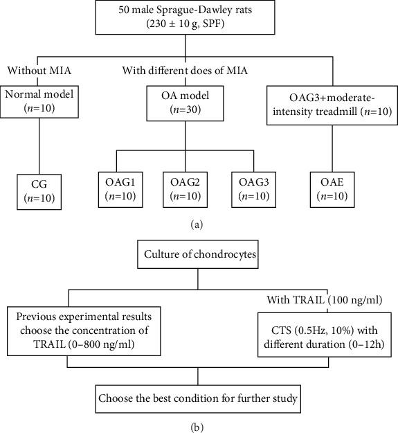
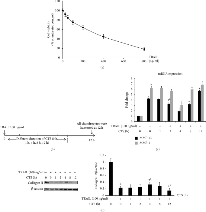
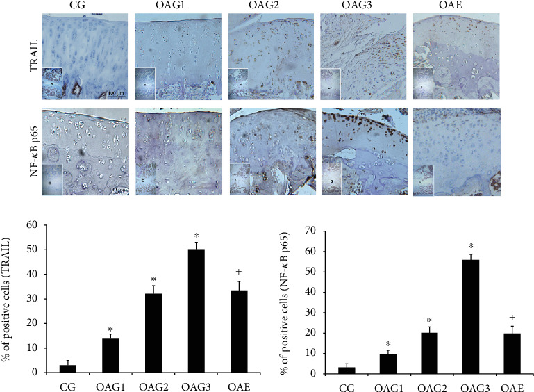
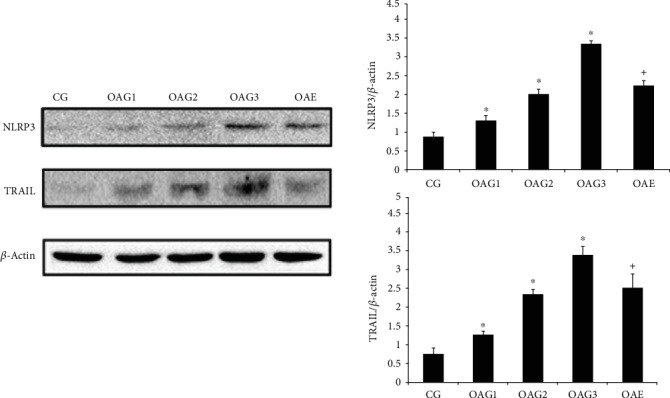
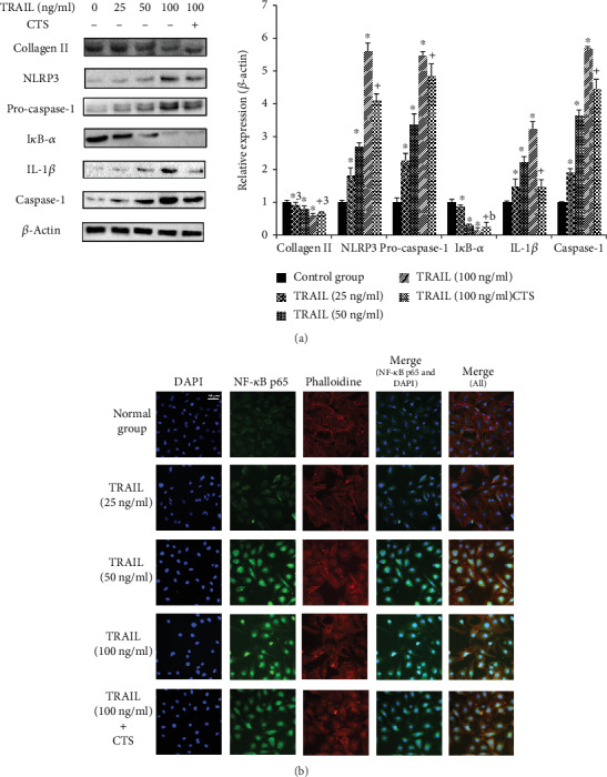
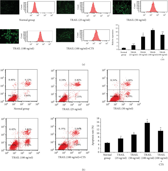
The aim of this study was to examine exercise-related genes in articular cartilage identified through bioinformatics analysis to dissect the potential signaling pathway involved in mechanical stimulation in osteoarthritis (OA). To this end, we evaluated the GSE74898 dataset from the Gene Expression Omnibus database for exercise-related differentially expressed miRNAs (DE-miRNAs) using the R software package and predicted potential target genes for these miRNAs using miRTarBase. Functional annotation and pathway enrichment analysis were performed for these potential DE-miRNA targets. The effects of mechanical stimulation on the tumor necrosis factor-related apoptosis-induced ligand (TRAIL)/nuclear factor-kappa B (NF-κB)/nucleotide-binding and oligomerization domain-like receptor containing protein 3 (NLRP3) signaling pathway were evaluated in articular cartilage and chondrocytes. A total of 394 DE-miRNAs were identified (103 upregulated miRNAs; 291 downregulated miRNAs) in the cartilage of rats following treadmill exercise compared to the cartilage of unexercised control rats. Thus, mechanical stimulation could modulate the TRAIL/NF-κB/NLRP3 signaling pathway on OA. Histological and protein analysis demonstrated that moderate-intensity treadmill exercise could ameliorate OA through the downregulation of TRAIL. Furthermore, moderate cyclic tensile strain (CTS) could rescue chondrocytes from the effects of TRAIL via the inhibition of the nuclear translocation of NF-κB p65 and formation of NLRP3. Our findings indicate that moderate mechanical stimulation could ameliorate the degeneration of cartilage and chondrocyte damage through the inhibition of the TRAIL/NF-κB/NLRP3 pathway.


Conflict of interest statement

The authors have declared that no competing interests exist.

Figures

Figure 1
Figure 1
The design of the treatment schedule. (a) The design of the animal experiment. Experimental groups: CG: control group; OAG1, OAG2, and OAG3: OA groups injected with different doses of MIA; OAE: OAG3 subjected to moderate-intensity treadmill exercise. (b) The design of the chondrocyte experiment. CTS: cyclic tensile strain; TRAIL: tumor necrosis factor-related apoptosis-induced ligand; MIA: monoiodoacetate; OA: osteoarthritis; SPF: specific-pathogen-free.
Figure 2
Figure 2
Western blot and qPCR analysis of chondrocytes treated with CTS for different durations. (a) The results of CCK-8 test. (b) The design of chondrocytes was subjected to CTS (10%, 0.5 Hz) for different durations (0 h, 1 h, 2 h, 4 h, 8 h, and 12 h) with TRAIL. (c) MMP-13 and MMP-1 mRNA expression in chondrocytes following different durations of CTS (10%, 0.5 Hz) was determined by qPCR. Differences between untreated and TRAIL (100 ng/ml)-induced chondrocytes (p < 0.001) and TRAIL-induced chondrocytes and those subjected to CTS for different durations (+p < 0.001, +ap = 0.001) were significant (ANOVA). Data are presented as the mean ± 95%confidence intervals; n = 9 per group. (d) Collagen II protein levels in chondrocytes. Differences between untreated and TRAIL (100 ng/ml)-induced chondrocytes (p < 0.001) and TRAIL-induced chondrocytes and those subjected to CTS of different durations (+p < 0.001, +ap = 0.006, and +bp = 0.002) were significant (ANOVA). β-Actin was used as an internal control. Data are presented as the mean ± 95%confidence intervals; n = 3 per group.
Figure 3
Figure 3
Immunohistochemical staining. The micrographs show the percentages of TRAIL-positive cells and the percentage of NF-κB p65 nuclear translocation in the articular cartilage of each experimental group. Differences between CG and OAG1, OAG2, and OAG3 (p < 0.001) and OAG3 and OAE (+p < 0.001) were significant (ANOVA). Data are presented as the mean ± 95%confidence intervals; n = 5 rats per group. Experimental groups: CG: control group; OAG1, OAG2, and OAG3: OA groups treated with different doses of MIA; OAE: OAG3 subjected to moderate-intensity treadmill exercise.
Figure 4
Figure 4
Protein expression levels were determined by western blotting of total protein extracted from cartilage. Differences between CG and OAG1, OAG2, and OAG3 (p < 0.001) and OAG3 and OAE (+p < 0.001) were significant (ANOVA). β-Actin was used as internal controls. Data are presented as the mean ± 95%confidence intervals; n = 3 rats per group. Experimental groups: CG: control group; OAG1, OAG2, and OAG3: OA groups treated with different doses of MIA; OAE: OAG3 subjected to moderate-intensity treadmill exercise.
Figure 5
Figure 5
Western blot and immunofluorescence analysis of chondrocytes. (a) Western blotting results for collagen II, NLRP3, procaspase-1, IκB-α, IL-1β, and caspase-1. Differences between untreated and TRAIL (0, 25, 50, and 100 ng/ml)-induced chondrocytes (p < 0.001, ∗ap = 0.005) and TRAIL (100 ng/ml)-induced chondrocytes and those subjected to CTS for 4 h (+p < 0.001, +ap = 0.003, and +bp = 0.001) were significant (ANOVA). Data are presented as the mean ± 95%confidence intervals; n = 3 per group. (b) Effects of CTS for 4 h on the nuclear translocation of NF-κB p65 in TRAIL-induced chondrocytes. The chondrocytes were immunostained using anti-NF-κB p65 rabbit antibody (green) and visualized by confocal microcopy. The cytoskeleton was visualized with phalloidin (red), and the cell nucleus was stained with 4,6-diamidino-2-phenylindole (DAPI; blue). Scale bar, 50 μm.
Figure 6
Figure 6
Reactive oxygen species (ROS) and apoptosis in chondrocytes. (a) Fluorescence microscopy and flow cytometry of ROS in chondrocytes. Differences between untreated and TRAIL (0, 25, 50, and 100 ng/ml)-induced chondrocytes (p < 0.001, ∗ap = 0.012) and TRAIL (100 ng/ml)-induced chondrocytes and those subjected to CTS for 4 h (+ap = 0.012) were significant (ANOVA). Data are presented as the mean ± 95%confidence intervals; n = 3 per group. (b) Chondrocyte apoptosis analysis by flow cytometry. Differences between untreated and TRAIL (0, 25, 50, and 100 ng/ml)-induced chondrocytes (p < 0.001) and TRAIL (100 ng/ml)-induced chondrocytes and those subjected to CTS for 4 h (+p < 0.001) were significant (ANOVA). Data are presented as the mean ± 95%confidence intervals; n = 3 in each group.

References

    1. Hunter D. J., Bierma-Zeinstra S. Osteoarthritis. The Lancet. 2019;393(10182):1745–1759. doi: 10.1016/S0140-6736(19)30417-9. - DOI - PubMed
    1. Ondrésik M., Azevedo Maia F. R., da Silva Morais A., et al. Management of knee osteoarthritis. Current status and future trends. Biotechnogy Bioengeering. 2017;114(4):717–739. doi: 10.1002/bit.26182. - DOI - PubMed
    1. Wallace I. J., Worthington S., Felson D. T., et al. Knee osteoarthritis has doubled in prevalence since the mid-20th century. Proceedings of the National Academy of Sciences of the United States of America. 2017;114(35):9332–9336. doi: 10.1073/pnas.1703856114. - DOI - PMC - PubMed
    1. Abbasi J. Can exercise prevent knee osteoarthritis? JAMA. 2017;318(22):2169–2171. doi: 10.1001/jama.2017.16144. - DOI - PubMed
    1. Emery C. A., Pasanen K. Current trends in sport injury prevention. Best Practice & Research Clinical Rheumatology. 2019;33(1):3–15. doi: 10.1016/j.berh.2019.02.009. - DOI - PubMed

MeSH terms