

Neuroprotective Effect of Chlorogenic Acid on Mitochondrial Dysfunction-Mediated Apoptotic Death of DA Neurons in a Parkinsonian Mouse Model
- PMID: 32566093
- PMCID: PMC7273475
- DOI: 10.1155/2020/6571484
Neuroprotective Effect of Chlorogenic Acid on Mitochondrial Dysfunction-Mediated Apoptotic Death of DA Neurons in a Parkinsonian Mouse Model
Abstract
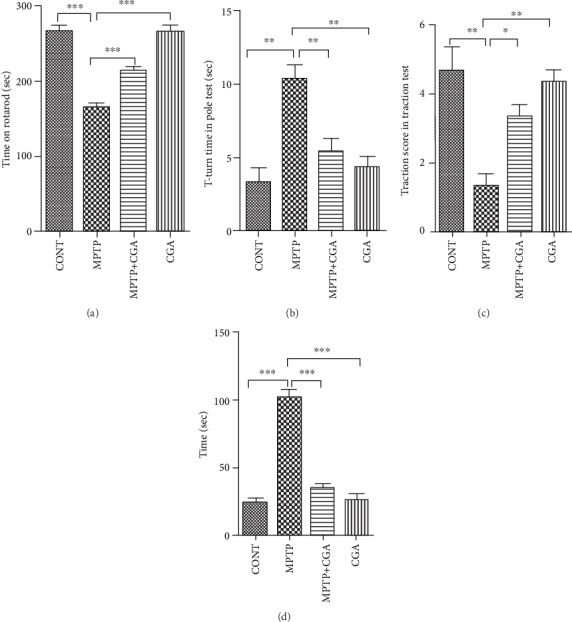
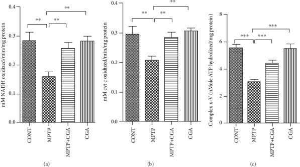
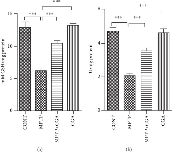
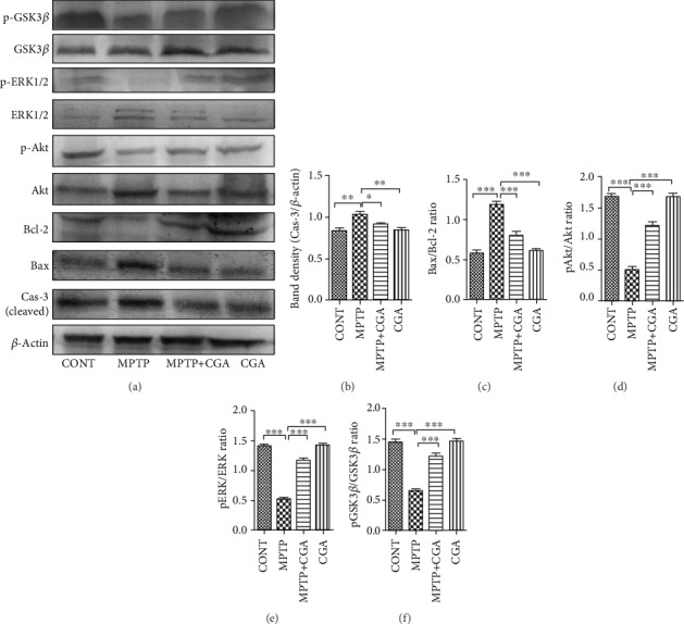
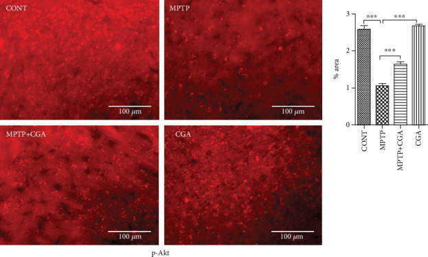
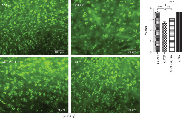
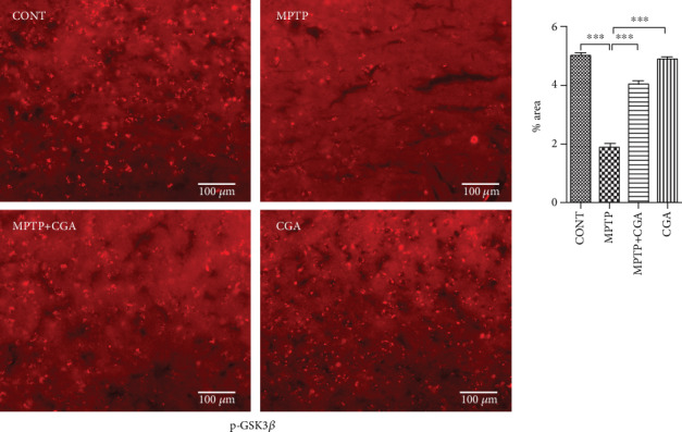
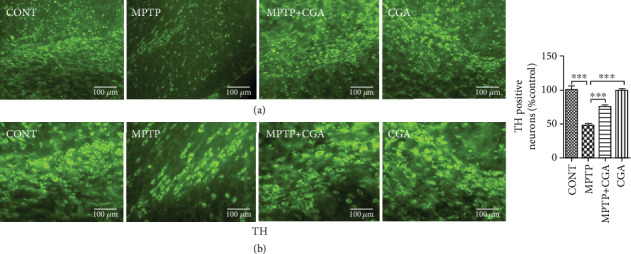
Mitochondrial dysfunction and oxidative stress characterize major factors involved in the activation of complex processes corresponding to apoptosis-mediated neuronal senescence of dopaminergic neurons (DA) in Parkinson's disease (PD). Here, we evaluated the molecular mechanisms participating in the treatment of a 1-methyl-4-phenyl-1,2,3,6-tetrahydopyridine- (MPTP-) intoxicated PD mouse model in response to chlorogenic acid (CGA). The results indicate that CGA treatment significantly improved the motor coordination of the MPTP-intoxicated mice. CGA also alleviated the fall in activity of mitochondrial complexes I, IV, and V in accordance with ameliorating the level of superoxide dismutase and mitochondrial glutathione in the midbrain of MPTP-induced mice. CGA inhibited the activation of proapoptotic proteins including Bax and caspase-3, while elevating the expression of antiapoptotic protein like Bcl-2 consequently preventing the MPTP-mediated apoptotic cascade. The study also revealed the improved phosphorylation state of Akt, ERK1/2, and GSK3β which was downregulated as an effect of MPTP toxicity. Our findings signify that CGA may possess pharmacological properties and contribute to neuroprotection against MPTP induced toxicity in a PD mouse model associated with phosphorylation of GSK3β via activating Akt/ERK signalling in the mitochondrial intrinsic apoptotic pathway. Thus, CGA treatment may arise as a potential therapeutic candidate for mitochondrial-mediated apoptotic senescence of DA neurons in PD.
Copyright © 2020 Saumitra Sen Singh et al.
Conflict of interest statement
All authors have no conflict of interest to report.
Figures
Similar articles
-
Neuroprotection by tetrahydroxystilbene glucoside in the MPTP mouse model of Parkinson's disease.Toxicol Lett. 2013 Oct 24;222(2):155-63. doi: 10.1016/j.toxlet.2013.07.020. Epub 2013 Aug 1. Toxicol Lett. 2013. PMID: 23911879
-
Neuroprotective properties of icariin in MPTP-induced mouse model of Parkinson's disease: Involvement of PI3K/Akt and MEK/ERK signaling pathways.Phytomedicine. 2017 Feb 15;25:93-99. doi: 10.1016/j.phymed.2016.12.017. Epub 2016 Dec 29. Phytomedicine. 2017. PMID: 28190476
-
Valproic Acid Protects Primary Dopamine Neurons from MPP+-Induced Neurotoxicity: Involvement of GSK3β Phosphorylation by Akt and ERK through the Mitochondrial Intrinsic Apoptotic Pathway.Biomed Res Int. 2017;2017:8124501. doi: 10.1155/2017/8124501. Epub 2017 Mar 22. Biomed Res Int. 2017. PMID: 28421199 Free PMC article.
-
MPTP-induced mouse model of Parkinson's disease: A promising direction of therapeutic strategies.Bosn J Basic Med Sci. 2021 Aug 1;21(4):422-433. doi: 10.17305/bjbms.2020.5181. Bosn J Basic Med Sci. 2021. PMID: 33357211 Free PMC article. Review.
-
Insights on the Correlation between Mitochondrial Dysfunction and the Progression of Parkinson's Disease.Endocr Metab Immune Disord Drug Targets. 2024;24(9):1007-1014. doi: 10.2174/0118715303249690231006114308. Endocr Metab Immune Disord Drug Targets. 2024. PMID: 37867265 Review.
Cited by
-
Oleuropein confers neuroprotection against rotenone-induced model of Parkinson's disease via BDNF/CREB/Akt pathway.Sci Rep. 2023 Feb 11;13(1):2452. doi: 10.1038/s41598-023-29287-4. Sci Rep. 2023. PMID: 36774383 Free PMC article.
-
Nanotechnology-Based Drug Delivery Strategies to Repair the Mitochondrial Function in Neuroinflammatory and Neurodegenerative Diseases.Pharmaceutics. 2021 Dec 1;13(12):2055. doi: 10.3390/pharmaceutics13122055. Pharmaceutics. 2021. PMID: 34959337 Free PMC article. Review.
-
WNT-β Catenin Signaling as a Potential Therapeutic Target for Neurodegenerative Diseases: Current Status and Future Perspective.Diseases. 2023 Jun 25;11(3):89. doi: 10.3390/diseases11030089. Diseases. 2023. PMID: 37489441 Free PMC article. Review.
-
Potential Applications of Mitochondrial Therapy with a Focus on Parkinson's Disease and Mitochondrial Transplantation.Adv Pharm Bull. 2024 Mar;14(1):147-160. doi: 10.34172/apb.2024.019. Epub 2023 Oct 14. Adv Pharm Bull. 2024. PMID: 38585467 Free PMC article. Review.
-
Chlorogenic Acid Improves PTSD-like Symptoms and Associated Mechanisms.Curr Neuropharmacol. 2021;19(12):2180-2187. doi: 10.2174/1570159X19666210111155110. Curr Neuropharmacol. 2021. PMID: 33430733 Free PMC article.
References
MeSH terms
Substances
LinkOut - more resources
Full Text Sources
Medical
Research Materials
Miscellaneous
