

The Rare Case of an Adult-Onset Xanthogranuloma of the Paranasal Sinuses: A Histological Dilemma
- PMID: 32566345
- PMCID: PMC7301186
- DOI: 10.1155/2020/2847821
The Rare Case of an Adult-Onset Xanthogranuloma of the Paranasal Sinuses: A Histological Dilemma
Abstract
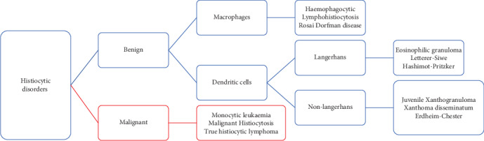
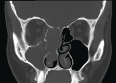
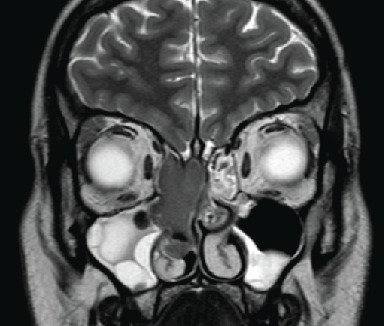
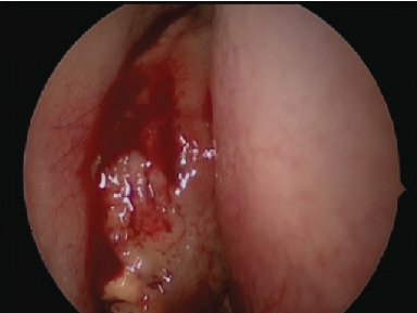
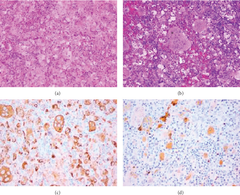
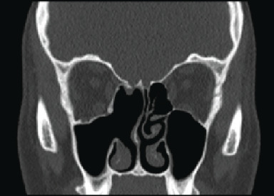
Xanthogranuloma is a rare benign tumour, part of the non-Langerhans cell histiocytosis group, uncommon in adults and even less common in the paranasal sinuses. Despite its benign nature, it mimics neoplasm due to its local effects which can have serious functional consequences depending on the anatomical location. We present the rare case of a young lady who presented insidiously with a maxillary sinus xanthogranuloma and was treated with endoscopic resection. Tissue diagnosis is of paramount importance to guide correct further investigations and management, and we discuss the potential challenge in identifying such a rarely seen pathology.
Copyright © 2020 J. Bastianpillai et al.
Conflict of interest statement
The authors declare that they have no conflicts of interest.
Figures
Similar articles
-
[Cladribine is highly effective in the treatment of Langerhans cell histiocytosis and rare histiocytic disorders of the juvenile xanthogranuloma group].Vnitr Lek. 2012 Jun;58(6):455-65. Vnitr Lek. 2012. PMID: 22913238 Review. Czech.
-
Xanthogranuloma formation after endoscopic sinus surgery: A case report.Int J Surg Case Rep. 2020;76:263-265. doi: 10.1016/j.ijscr.2020.09.200. Epub 2020 Oct 2. Int J Surg Case Rep. 2020. PMID: 33053486 Free PMC article.
-
Unilateral pathological lesions of paranasal sinuses removed by endoscopic surgery.Otolaryngol Pol. 2014 Mar-Apr;68(2):83-8. doi: 10.1016/j.otpol.2013.07.002. Epub 2013 Jul 5. Otolaryngol Pol. 2014. PMID: 24629740
-
[Distant metastases to nasal cavities and paranasal sinuses, from the organs outside the head and neck].Otolaryngol Pol. 2008;62(4):422-5. doi: 10.1016/S0030-6657(08)70284-1. Otolaryngol Pol. 2008. PMID: 18837216 Polish.
-
Adenoid cystic carcinoma of the paranasal sinuses: retrospective series and review of the literature.Eur Ann Otorhinolaryngol Head Neck Dis. 2013 Nov;130(5):257-62. doi: 10.1016/j.anorl.2012.09.010. Epub 2013 Jun 6. Eur Ann Otorhinolaryngol Head Neck Dis. 2013. PMID: 23747147 Review.
References
-
- Avelino M. A., Elias T. G. A., Rezende R. M. D., Lima A. P. L., Gonçalves A. V., Gonçalves A. V. Rosai-Dorfman em seios paranasais como diagnóstico diferencial de polipose nasosinusal na infância. Brazilian Journal of Otorhinolaryngology. 2012;78(3):136–136. doi: 10.1590/S1808-86942012000300024. - DOI - PMC - PubMed
Publication types
LinkOut - more resources
Full Text Sources
