Review
. 2020 Apr;30(Suppl 1):S45-S53.
doi: 10.4103/jcecho.jcecho_7_19. Epub 2020 Apr 10.

Cardiac Tumors

Affiliations
Review

Cardiac Tumors

Grazia Casavecchia et al. J Cardiovasc Echogr. 2020 Apr.

Abstract

Cardiac tumors (CTs) are extremely rare, with an incidence of approximately 0.02% in autopsy series. Primary tumors of the heart are far less common than metastatic tumors. CTs usually present with any possible clinical combination of heart failure, arrhythmias, or embolism. Echocardiography remains the first diagnostic approach when suspecting a CT which, on the other side, frequently appears unexpectedly during an echocardiographic examination. Yet, cardiac tomography and especially magnetic resonance imaging may offer several adjunctive opportunities in the diagnosis of CTs. Early and exact diagnosis is crucial for the following therapy and outcome of CTs.

Keywords: Atrial myxomas; cancer of the heart; cardiac tumors; computed tomography and magnetic resonance imaging of primary cardiac malignancies; echocardiography in the diagnosis of cardiac tumors; malignant tumors of the heart.


Conflict of interest statement

There are no conflicts of interest.

Figures
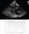
Figure 1
Figure 1
Metastatic angiosarcoma infiltrating the right ventricular wall. Top: 2D-echo long axis view. Down: pseudoischemic anomalies on corresponding leads
Figure 2
Figure 2
Papillary Fibroelastoma, 67-year old patient after transient ischemic attack (30 days earlier). 2D (left) and 3D (right) transthoracic echocardiogram showing a round formation with soft consistence on the atrial side of septal leaflet of tricuspid valve, firstly and erroneously considered ad an endocardial vegetation
Figure 3
Figure 3
(a) 2D apical 3-chamber view; (b) 2D apical 4-chamber view. Pericardial effusion and infiltrative mass on left and right ventricular apex, suggestive for malignancy in hip liposarcoma, endometrial adenocarcinoma, and breast infiltrating carcinoma. Final diagnosis was lymphoma
Figure 4
Figure 4
Echocardiogram of cardiac lymphoma infiltrating the left ventricular wall and protruding into the left ventricular lumen, after contrast injection. The apical septum is partially spared from neoplastic infiltration and appears darker. The small residual left ventricular cavity appears brighter (contrast effect
Figure 5
Figure 5
TEE of right atrial angiosarcoma extendig to superior vena cava. 2D (left) and 3D (right) images (Courtesy of Dr. Rita Piazza)
Figure 6
Figure 6
Transvenous biopsy of right atrial angiosarcoma (see figure 5). TEE monitoring was used to assess the position of the bioptome (arrow) (Courtesy of Dr. Rita Piazza)
Figure 7
Figure 7
cRMI: left, iso-intense in T1-TSE sequences; middle iper-intense in T2; right, PSIR with central hypo-intense core compatible with diagnosis of fibroelastoma (courtesy Dr. Matteo Gravina)
Figure 8
Figure 8
Lypoma, 60-year old woman, with poor acustic window echocardiogram. Up, cMRI showing rounded capsule formation in right atrium on interatrial septum. Down, hyper-intense in T1 sequence and hypo-intense in fat sat sequences compatible with lypoma (courtesy of Dr. Matteo Gravina)
Figure 9
Figure 9
Cardiac Fibroma, 51-year old man referred for dizziness, sweating and vomit, admitted to acute cardiac care unit with suspected acute coronary syndrome (a) Echocardiography apical 4-chamber view: the mass is not visible. (b) Echocardiography off axis view: oval mass of the left ventricular lateral wall (ref 3940)
Figure 10
Figure 10
Cardiac fibroma. Upper, cMRI T2-weighted images 4-chamber and short axis view showing hypo-intense nodular mass in the mid-apical and inferior-lateral wall segments. Down-left, first-pass minimal enhancement with gadolinium. Down-right, late enhancement 4-chamber view, the mass appears as hyper-intense (ref 3940)
Figure 11
Figure 11
Angiosarcoma, 68-year old male with history of chest pain and dyspnea. cMRI: Solid tissue with improved contrast enhancement, surrounding the superior vena cava and right atrium and leading to reduced expansion with pericardium thickening and minimal effusion (courtesy of Dr. Matteo Gravina)

References

    1. Neragi-Miandoab S, Kim J, Vlahakes GJ. Malignant tumours of the heart: A review of tumour type, diagnosis and therapy. Clin Oncol (R Coll Radiol) 2007;19:748–56. - PubMed
    1. Rizzo S, Thiene G, Valente M, Basso C. Primary cardiac malignancies: Epidemiology and pathology. In: Lestuzzi C, Oliva S, Ferraù F, editors. Manual of Cardioncology. Springer; 2017. pp. 339–65.
    1. Sarjeant JM, Butany J, Cusimano RJ. Cancer of the heart: Epidemiology and management of primary tumors and metastases. Am J Cardiovasc Drugs. 2003;3:407–21. - PubMed
    1. Pino P, Lestuzzi C. Cardiac malignancies. Clinical aspects. In: Lestuzzi C, Oliva S, Ferraù F, editors. Manual of Cardioncology. Springer; 2017. p. 311-.
    1. Lestuzzi C, Nicolosi GL, Biasi S, Piotti P, Zanuttini D. Sensitivity and specificity of electrocardiographic ST-T changes as markers of neoplastic myocardial infiltration. Echocardiographic correlation. Chest. 1989;95:980–5. - PubMed