

Acute disseminated encephalomyelitis after dengue
- PMID: 32566482
- PMCID: PMC7298550
- DOI: 10.1016/j.idcr.2020.e00862
Acute disseminated encephalomyelitis after dengue
Abstract
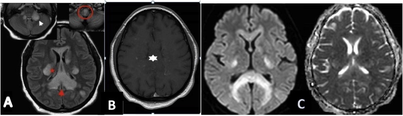
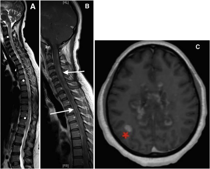
Dengue fever, transmitted by Aedes aegypti mosquitoes, is one of the most common vector-borne disease. Its incidence is increasing steadily worldwide, becoming a major public health problem in the tropical and subtropical zone. Neurological manifestations after dengue are not very common and acute disseminated encephalomyelitis (ADEM) following dengue infections is rare with a few cases documented in literature. Clinical characteristics and typical lesions of ADEM on magnetic resonance imaging (MRI) of brain along with serologic positivity for dengue usually confirm the diagnosis. We report a case of ADEM which developed as a neurological complication of dengue during an epidemic in a 39-year-old woman.
Keywords: ADEM, Acute Disseminated Encephalomyelitis; Acute disseminated encephalomyelitis; CSF, Cerebrospinal Fluid; Dengue infection; EEG, Electroencephalography; MRI, Magnetic Resonance Imaging; Mayotte; PCR, Polymerase Chain Reaction.
© 2020 The Authors.
Figures
References
-
- Sinniah M., Igarashi A. Dengue haemorrhagic fever. Rev Med Virol. 1995;5:193–203.
-
- Murthy J.M. Neurological complication of dengue infection. Neurol India. 2010;58:581–584. - PubMed
-
- Sanjeev Kumar B., Naik S., Jayantee K., Misra U.K. Acute disseminated encephalomyelitis following dengue virus infection. J Neuroinfect Dis. 2014;5:139. doi: 10.4172/2314-7326.1000139. - DOI
-
- Gera C., George U. Acute disseminating encephalomyelitis with haemorrhage following dengue. Neurol India. 2010;58:595–596. - PubMed
Publication types
LinkOut - more resources
Full Text Sources
