. 2020 May;8(10):637.
doi: 10.21037/atm-20-2119.

The epidemiologic and clinical features of suspected and confirmed cases of imported 2019 novel coronavirus pneumonia in north Shanghai, China

Affiliations

The epidemiologic and clinical features of suspected and confirmed cases of imported 2019 novel coronavirus pneumonia in north Shanghai, China

Shuanshuan Xie et al. Ann Transl Med. 2020 May.

Abstract

Background: A recent cluster of pneumonia cases in Wuhan (China) is known to be caused by a novel beta-coronavirus named the corona virus disease 2019 (COVID-19) and can be spread through human-to-human transmission.

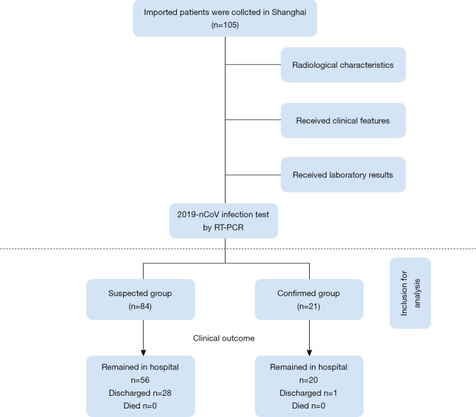
Methods: Data of 21 patients with laboratory-confirmed COVID-19 and 84 patients with suspected COVID-19 were analyzed by RT-PCR. The epidemiologic and clinical features as well as clinical outcomes were compared between the confirmed and suspected cases.

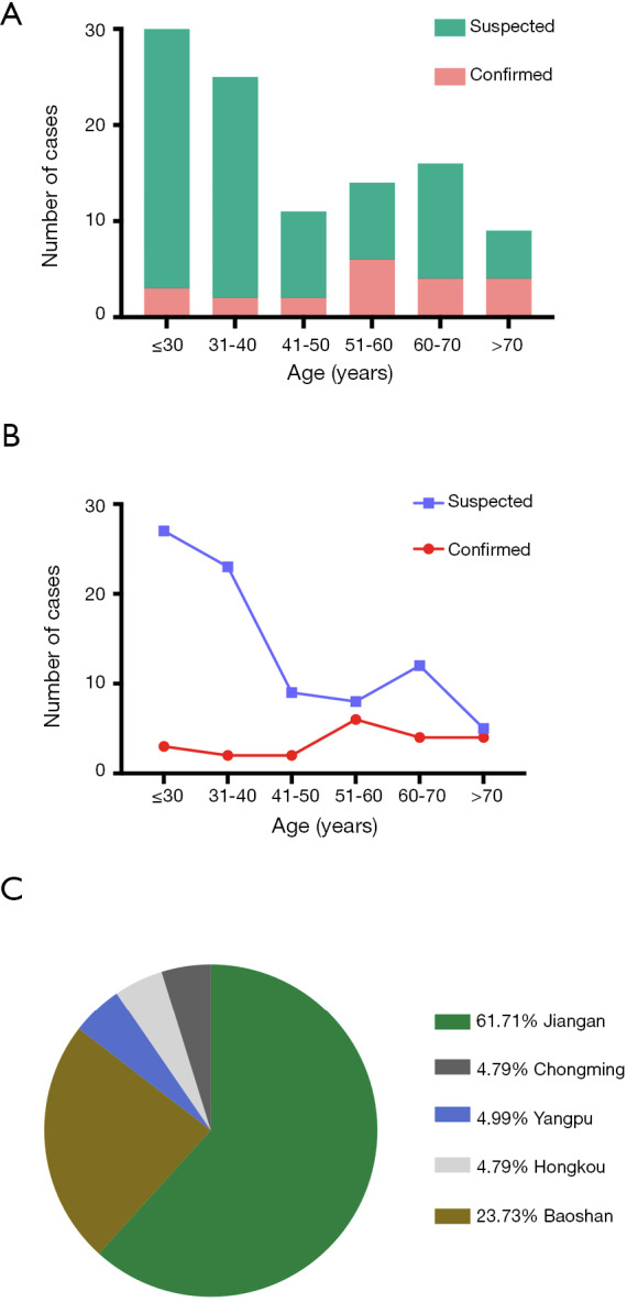
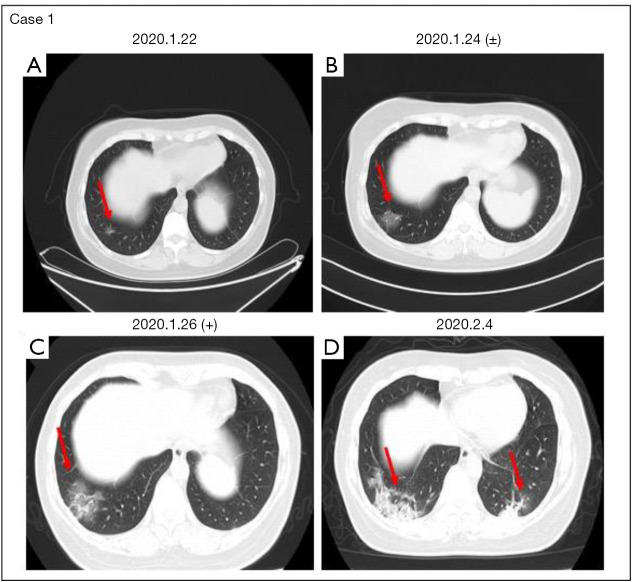
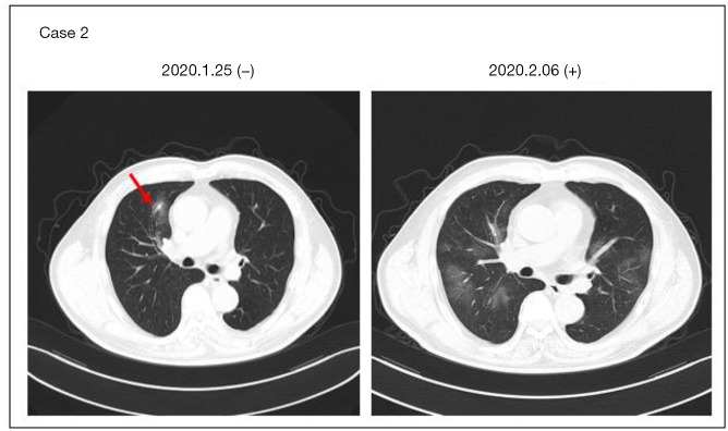
Results: Altogether 105 patients had been enrolled in this study by February 15, 2020 in north Shanghai, including 21 confirmed cases and 84 suspected cases of COVID-19. The incubation period of these confirmed patients with imported COVID-19 was 17.6 days (IQR 5-34 days) and the median time from symptom onset to diagnosis was 145.64 h (IQR 21-441 h). More than 50% of the confirmed patients were older than 51 (range, 51-60) years. Fifty (59.5%) of the 84 probably patients were younger than 40 years, including 27 (32%) patients younger than 30 years. Most confirmed patients were men (61.9%, 13/21), and less than 50% of them had underlying diseases, including diabetes (9.5%, 2/21), hypertension (19%, 4/21), COPD (23.8%, 5/21), and CD (23.8%, 5/21). In addition, 10 (47.6%) of the 21 confirmed patients were ordinary employees, and 12 (57.2%) of them had recently been to Wuhan or had close contacts with people from Wuhan. Of the 84 suspected patients, 28 (33.3%) were retired employees; 69 (82.1%) had recently been to supermarkets and groceries or had a history of traveling abroad or to other cities of China. The common onset symptoms of the patients in both groups were fever and cough. The symptom of Sputum production was more pronounced in probably patients (40.5%, 34/84) than that in confirmed patients (9.5%, 2/21). More than 50% imported patients (53.3%, 56/105) had one and two affected lobes. Twenty-nine (27.6%) of the 105 imported patients had been discharged, no patient had died, and all the other patients are still in hospital.

Conclusions: The overall incubation period in this cohort of imported confirmed COVID-19 patients was longer than that in Wuhan, mostly infecting older men. The disease onset of imported COVID-19 infection was occult, and the clinical symptoms were usually mild, mostly presenting as low fever, fatigue, light cough, and mild dyspnea.

Keywords: Coronavirus infections; computed tomographic findings; imported; incubation period; pneumonia; symptoms.


Conflict of interest statement

Conflicts of Interest: All authors have completed the ICMJE uniform disclosure form (available at http://dx.doi.org/10.21037/atm-20-2119). The authors have no conflicts of interest to declare.

Figures

Figure 1
Figure 1
Flow chart of patient selection for the study.
Figure 2
Figure 2
Age and residence distributions of the present study. (A,B) Bar charts and line chart demonstrating the number of cases (y-axis) and age distribution (x-axis) of confirmed and suspected cases. (C) Pie charts demonstrating the etiology of residence based on retrospective diagnosis in the confirmed group.
Figure 3
Figure 3
Case 1 in the confirmed group. Case 1: chest CT and PCR demonstrating progression from weak positivity to positivity. (A) A small inflamed nodule is seen in the right lower lobe of the lung, and no obvious exudation is seen in other lung fields. (B) A flocculent fuzzy exudation shadow is seen in the basal segment of the right inferior lung lobe. At the same time, RT-PCR shows a weakly positive result of COVID-19. (C) CT shows a worse status: a large GGO is seen in the lower right lobe. Tracheal intubation is seen in the trachea. COVID-19 is positive by RT-PCR. Chest CT on 2020-02-04 (D) shows high-density shadows in both lungs. GGO, ground-glass opacity. The red arrows indicate lesion location.
Figure 4
Figure 4
Case 2 in the confirmed group. Case 2: chest CT and two consecutive RT-PCR assays for 2COVID-19 demonstrating progression from negativity to positivity. Chest CT on 2020-01-25 (A) shows exudation and a nodular shadow near the bronchus of the upper lobe of the right lung, and two consecutive RT-PCR assays for COVID-19 on 01-25 and 01-26 are both negative. Chest CT on 2020-02-06 (B) shows multiple GGO exudation shadows in both lungs. COVID-19 is positive and chest CT shows a worse status. GGO, ground-glass opacity. The red arrow indicates lesion location.
Figure 5
Figure 5
Chest CT of two suspected cases. Case 1: chest CT shows high-density mass shadows in the right lungs (A). After one week, chest CT shows no significant change (B). Case 2: CT shows a density shadow of mottled GGO in the right lobe (C). After short-term treatment, chest CT shows significant changes (D). GGO, ground-glass opacity. The red arrows indicate lesion location.
Figure S1
Figure S1
Chest CT of 10 confirmed cases before and after treatment. Cases 3, 4, 5: chest CT (left) shows high-density mass shadows in both lungs. Tracheal intubation is seen in the trachea. Chest CT shows an improved status (right). Case 6: CT shows density shadows of mottled GGO in the right lung. Chest CT shows a worse status (right). Case 8: CT shows density shadows of mottled GGO in the left lung. Chest CT shows an improved status (right). Cases 9, 10: chest CT (left) shows density shadows of flake GGO in both lungs. Chest CT shows an improved status (right). Case 11: CT shows density shadows of mottled GGO in the left lung. Chest CT shows a worse status (right). Case 7: chest CT shows a worse status (right). CT shows density shadows of mottled GGO in the left lung. Case 12: chest CT shows nodular consolidation with partial GGO in the right lung. chest CT shows no change (right). GGO, ground-glass opacity.

References

    1. Lu H, Stratton CW, Tang YW. Outbreak of pneumonia of unknown etiology in Wuhan China: the mystery and the miracle. J Med Virol 2020;92:401-2. 10.1002/jmv.25678 - DOI - PMC - PubMed
    1. Hui DS, I, Azhar E, Madani TA, et al. The continuing 2019nCoV epidemic threat of novel coronaviruses to global health the latest 2019 novel coronavirus outbreak in Wuhan, China. Int J Infect Dis 2020;91:264-6. 10.1016/j.ijid.2020.01.009 - DOI - PMC - PubMed
    1. Huang C, Wang Y, Li X, et al. Clinical features of patients infected with 2019 novel coronavirus in Wuhan, China. Lancet 2020;395:497-506 10.1016/S0140-6736(20)30183-5 - DOI - PMC - PubMed
    1. WHO. Novel coronavirus–China. Jan 12, 2020. Available online: http://www.who.int/csr/don/12-january-2020-novel-coronavirus-china/en/ (accessed Jan 19, 2020).
    1. WHO. Novel coronavirus–Thailand (ex-China). Jan 14, 2020. http://www.who.int/csr/don/14-january-2020-novel-coronavirus-thailand/en/ (accessed Jan 19, 2020).