Review
. 2020 May 22:2020:7608964.
doi: 10.1155/2020/7608964. eCollection 2020.

Pathophysiology and Management of Type 2 Diabetes Mellitus Bone Fragility

Affiliations
Review

Pathophysiology and Management of Type 2 Diabetes Mellitus Bone Fragility

C Eller-Vainicher et al. J Diabetes Res. .

Abstract

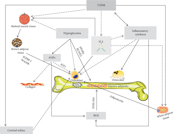
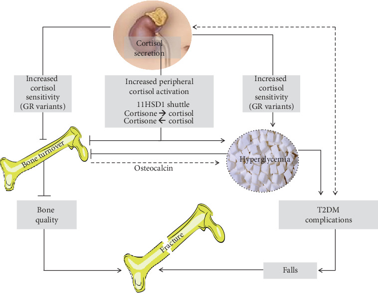
Individuals with type 2 diabetes mellitus (T2DM) have an increased risk of bone fragility fractures compared to nondiabetic subjects. This increased fracture risk may occur despite normal or even increased values of bone mineral density (BMD), and poor bone quality is suggested to contribute to skeletal fragility in this population. These concepts explain why the only evaluation of BMD could not be considered an adequate tool for evaluating the risk of fracture in the individual T2DM patient. Unfortunately, nowadays, the bone quality could not be reliably evaluated in the routine clinical practice. On the other hand, getting further insight on the pathogenesis of T2DM-related bone fragility could consent to ameliorate both the detection of the patients at risk for fracture and their appropriate treatment. The pathophysiological mechanisms underlying the increased risk of fragility fractures in a T2DM population are complex. Indeed, in T2DM, bone health is negatively affected by several factors, such as inflammatory cytokines, muscle-derived hormones, incretins, hydrogen sulfide (H2S) production and cortisol secretion, peripheral activation, and sensitivity. All these factors may alter bone formation and resorption, collagen formation, and bone marrow adiposity, ultimately leading to reduced bone strength. Additional factors such as hypoglycemia and the consequent increased propensity for falls and the direct effects on bone and mineral metabolism of certain antidiabetic medications may contribute to the increased fracture risk in this population. The purpose of this review is to summarize the literature evidence that faces the pathophysiological mechanisms underlying bone fragility in T2DM patients.


Conflict of interest statement

The authors declare that they have no competing interests.

Figures

Figure 1
Figure 1
PRISMA flow diagram. According to PRISMA guidelines, PubMed and MEDLINE were searched from June 1968 to January 2020 for identifying published articles about bone metabolism and T2DM. In particular, we considered articles focused on the interactions between T2DM and bone fragility, such as hyperglycemia, insulin resistance, AGEs, bone marrow adiposity, inflammatory cytokines, H2S, and cortisol. Studies that analyzed how T2DM impacts on bone formation and resorption, collagen formation, skeletal muscle, and the incretin system were evaluated. Only publications in English were included.
Figure 2
Figure 2
Mechanisms underlying bone fragility in type 2 diabetes mellitus (T2DM). In T2DM, the muscle tissue reduction, due to several factors including hyperglycemia per se, but probably also hydrogen sulfide (H2S) decrease, is thought to have a negative role on osteoblast lineage, via its crosstalk with the brown adipose tissue. Indeed, the muscle tissue is known to influence the brown adipose tissue, physiologically stimulating the secretion of factors (such as IGFBP2 and Wnt10b) thought to be important for osteoblast proliferation and activity. Osteoblast differentiation and activity, in T2DM, may be also impaired directly by the reduction of H2S levels that physiologically are thought to stimulate the osteoblast lineage. Hyperglycemia may directly reduce bone mesenchymal stem cell (MSC) viability and clonogenicity and also have an indirect negative effect on osteoblasts via the accumulation of advanced glycation end products (AGEs), which negatively affects osteoblasts through a reduction of the insulin-like growth factor-1 (IGF1) levels. The AGE accumulation impairs the normal collagen formation and leads to reactive oxygen species (ROS) increase that may augment marrow adiposity via the phosphoinositide-3-kinase–protein kinase B/Akt (PI3K/Akt) pathway. The inflammatory cytokine increase, directly and/or indirectly (due to the H2S reduction), may also impair osteoblastogenesis and increase osteoclast activity and ROS, ultimately leading to bone adiposity. Finally, in T2DM, osteoblasts may be also damaged by the low adiponectin levels due to the increase of white adipose tissue, which is a characteristic of T2DM itself but also a consequence of low H2S levels. Finally, even an altered cortisol secretion, peripheral activation, and sensitivity (i.e., “cortisol milieu”) have been suggested to potentially impair osteoblast activity.
Figure 3
Figure 3
Cortisol milieu and bone fragility in type 2 diabetes mellitus (T2DM). In T2DM patients, an increased (even though still within the normal range) cortisol secretion is present, particularly in those affected with the diabetic complications, which in turn is hypothesized to be a trigger for the increased cortisol secretion itself. The sensitizing variants of the glucocorticoid receptor (GR) may increase the negative effect of cortisol on both T2DM control and bone metabolism, contributing to the shift in the balance between osteoblastogenesis and adipogenesis of mesenchymal stem cells in bone. The degree of the interconversion of cortisone in cortisol, due to the activity of the 11βhydroxysteroidodehydrogenase type 1 (11HSD1), may influence bone metabolism in T2DM. Indeed, in humans, the selective inhibition of 11HSD1, which has been even suggested as potential treatment for T2DM, has been also demonstrated to improve diabesity and osteoblast differentiation in a mouse model. Finally, in T2DM, a vicious circle could be hypothesized between the increased cortisol secretion, peripheral activation, and sensitivity (i.e., “cortisol milieu”) and bone and glycometabolic control. Indeed, the low bone turnover induced by this activated cortisol milieu could contribute in reducing the undercarboxylated osteocalcin levels, which decrease and, in turn, may worsen the glycometabolic control, therefore perpetuating the mechanisms leading to reduced bone turnover. The final effects of these alterations of the cortisol milieu in T2DM may be on one side of the reduction of bone quality, since the low bone turnover reduces the possibility of the microcrack repairing, and, on the other side, the worsening of the T2DM complications that ultimately could lead to an increased risk of falls. The reduction of bone quality together with the increased risk of falls is among the most important factors associated with bone fragility in T2DM.

References

    1. Valderrábano R. J., Linares M. I. Diabetes mellitus and bone health: epidemiology, etiology and implications for fracture risk stratification. Clinical Diabetes and Endocrinology. 2018;4(1) doi: 10.1186/s40842-018-0060-9. - DOI - PMC - PubMed
    1. Napoli N., Chandran M., Pierroz D. D., Abrahamsen B., Schwartz A. V., Ferrari S. L. Mechanisms of diabetes mellitus-induced bone fragility. Mechanisms of diabetes mellitus-induced bone fragility. Nature Reviews. Endocrinology. 2017;13(4):208–219. doi: 10.1038/nrendo.2016.153. - DOI - PubMed
    1. Vestergaard P. Discrepancies in bone mineral density and fracture risk in patients with type 1 and type 2 diabetes—a meta-analysis. Osteoporosis International. 2007;18(4):427–444. doi: 10.1007/s00198-006-0253-4. - DOI - PubMed
    1. Janghorbani M., van Dam R. M., Willett W. C., Hu F. B. Systematic review of type 1 and type 2 diabetes mellitus and risk of fracture. American Journal of Epidemiology. 2007;166(5):495–505. doi: 10.1093/aje/kwm106. - DOI - PubMed
    1. Fan Y., Wei F., Lang Y., Liu Y. Diabetes mellitus and risk of hip fractures: a meta-analysis. Osteoporosis International. 2016;27(1):219–228. doi: 10.1007/s00198-015-3279-7. - DOI - PubMed

MeSH terms

Substances